

127
1. Prepare solutions with varying concentrations (2–10,000 nM)
of LH 2 -AMP in MOPS buffer.
2. Follow the steps 2–4 in Subheading 3.6.
1. Suspend equimolar BD1/Donor and BD2/Acceptor (20–
500 nM) with varying concentrations of rapamycin in the
MOPS buffer (see Note 3).
2. Dispense the mixture (50 μL) to an assay tube for a
luminometer.
3. Measure the light intensity after injecting 450 μL of 1.1 mM
ATP, 83 μM of LH 2 , and 1.1 mM coenzyme A with 10-msec
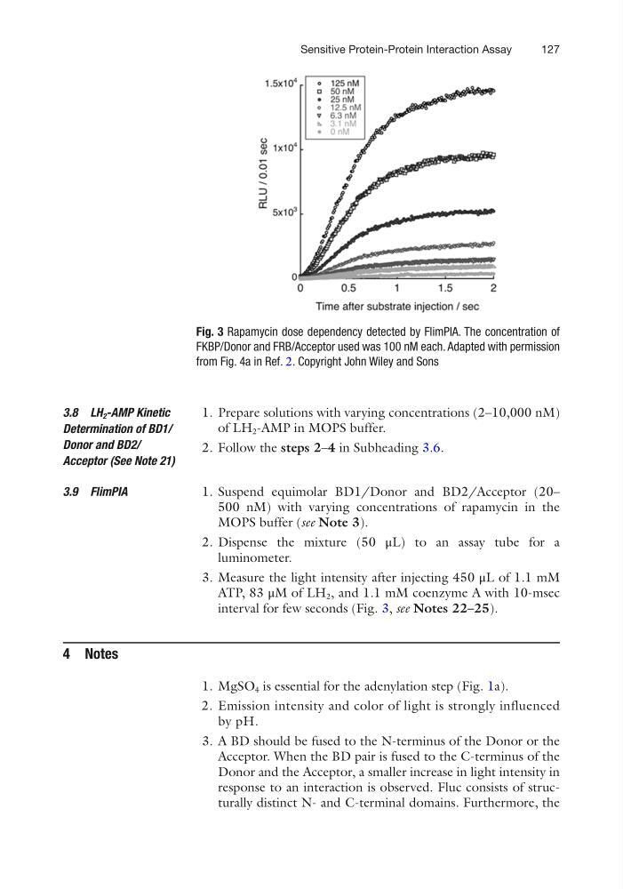
interval for few seconds (Fig. 3, see Notes 22–25).
4 Notes
1. MgSO 4 is essential for the adenylation step (Fig. 1a).
2. Emission intensity and color of light is strongly influenced
by pH.
3. A BD should be fused to the N-terminus of the Donor or the
Acceptor. When the BD pair is fused to the C-terminus of the
Donor and the Acceptor, a smaller increase in light intensity in
response to an interaction is observed. Fluc consists of structurally distinct N- and C-terminal domains. Furthermore, the
3.8 LH 2 -AMP Kinetic
Determination of BD1/
Donor and BD2/
Acceptor (See Note 21)
3.9 FlimPIA
Fig. 3 Rapamycin dose dependency detected by FlimPIA. The concentration of
FKBP/Donor and FRB/Acceptor used was 100 nM each. Adapted with permission
from Fig. 4a in Ref. 2. Copyright John Wiley and Sons
Sensitive Protein-Protein Interaction Assay
1. Prepare solutions with varying concentrations (2–10,000 nM)
of LH 2 -AMP in MOPS buffer.
2. Follow the steps 2–4 in Subheading 3.6.
1. Suspend equimolar BD1/Donor and BD2/Acceptor (20–
500 nM) with varying concentrations of rapamycin in the
MOPS buffer (see Note 3).
2. Dispense the mixture (50 μL) to an assay tube for a
luminometer.
3. Measure the light intensity after injecting 450 μL of 1.1 mM
ATP, 83 μM of LH 2 , and 1.1 mM coenzyme A with 10-msec
interval for few seconds (Fig. 3, see Notes 22–25).
4 Notes
1. MgSO 4 is essential for the adenylation step (Fig. 1a).
2. Emission intensity and color of light is strongly influenced
by pH.
3. A BD should be fused to the N-terminus of the Donor or the
Acceptor. When the BD pair is fused to the C-terminus of the
Donor and the Acceptor, a smaller increase in light intensity in
response to an interaction is observed. Fluc consists of structurally distinct N- and C-terminal domains. Furthermore, the
3.8 LH 2 -AMP Kinetic
Determination of BD1/
Donor and BD2/
Acceptor (See Note 21)
3.9 FlimPIA
Fig. 3 Rapamycin dose dependency detected by FlimPIA. The concentration of
FKBP/Donor and FRB/Acceptor used was 100 nM each. Adapted with permission
from Fig. 4a in Ref. 2. Copyright John Wiley and Sons
Sensitive Protein-Protein Interaction Assay