

82
H. Zhu et al.
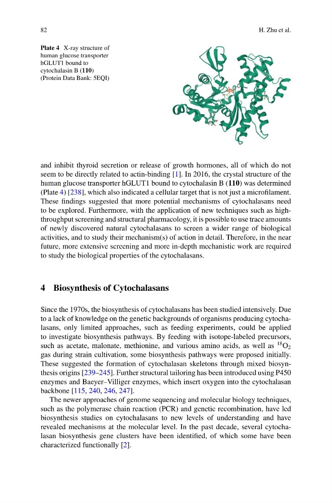
Plate 4 X-ray structure of
human glucose transporter
hGLUT1 bound to
cytochalasin B (110)
(Protein Data Bank: 5EQI)
and inhibit thyroid secretion or release of growth hormones, all of which do not
seem to be directly related to actin-binding [1]. In 2016, the crystal structure of the
human glucose transporter hGLUT1 bound to cytochalasin B (110) was determined
(Plate 4) [238], which also indicated a cellular target that is not just a microfilament.
These findings suggested that more potential mechanisms of cytochalasans need
to be explored. Furthermore, with the application of new techniques such as highthroughput screening and structural pharmacology, it is possible to use trace amounts
of newly discovered natural cytochalasans to screen a wider range of biological
activities, and to study their mechanism(s) of action in detail. Therefore, in the near
future, more extensive screening and more in-depth mechanistic work are required
to study the biological properties of the cytochalasans.
4 Biosynthesis of Cytochalasans
Since the 1970s, the biosynthesis of cytochalasans has been studied intensively. Due
to a lack of knowledge on the genetic backgrounds of organisms producing cytochalasans, only limited approaches, such as feeding experiments, could be applied
to investigate biosynthesis pathways. By feeding with isotope-labeled precursors,
such as acetate, malonate, methionine, and various amino acids, as well as
18 O 2
gas during strain cultivation, some biosynthesis pathways were proposed initially.
These suggested the formation of cytochalasan skeletons through mixed biosynthesis origins [239–245]. Further structural tailoring has been introduced using P450
enzymes and Baeyer–Villiger enzymes, which insert oxygen into the cytochalasan
backbone [115, 240, 246, 247].
The newer approaches of genome sequencing and molecular biology techniques,
such as the polymerase chain reaction (PCR) and genetic recombination, have led
biosynthesis studies on cytochalasans to new levels of understanding and have
revealed mechanisms at the molecular level. In the past decade, several cytochalasan biosynthesis gene clusters have been identified, of which some have been
characterized functionally [2].
H. Zhu et al.
Plate 4 X-ray structure of
human glucose transporter
hGLUT1 bound to
cytochalasin B (110)
(Protein Data Bank: 5EQI)
and inhibit thyroid secretion or release of growth hormones, all of which do not
seem to be directly related to actin-binding [1]. In 2016, the crystal structure of the
human glucose transporter hGLUT1 bound to cytochalasin B (110) was determined
(Plate 4) [238], which also indicated a cellular target that is not just a microfilament.
These findings suggested that more potential mechanisms of cytochalasans need
to be explored. Furthermore, with the application of new techniques such as highthroughput screening and structural pharmacology, it is possible to use trace amounts
of newly discovered natural cytochalasans to screen a wider range of biological
activities, and to study their mechanism(s) of action in detail. Therefore, in the near
future, more extensive screening and more in-depth mechanistic work are required
to study the biological properties of the cytochalasans.
4 Biosynthesis of Cytochalasans
Since the 1970s, the biosynthesis of cytochalasans has been studied intensively. Due
to a lack of knowledge on the genetic backgrounds of organisms producing cytochalasans, only limited approaches, such as feeding experiments, could be applied
to investigate biosynthesis pathways. By feeding with isotope-labeled precursors,
such as acetate, malonate, methionine, and various amino acids, as well as
18 O 2
gas during strain cultivation, some biosynthesis pathways were proposed initially.
These suggested the formation of cytochalasan skeletons through mixed biosynthesis origins [239–245]. Further structural tailoring has been introduced using P450
enzymes and Baeyer–Villiger enzymes, which insert oxygen into the cytochalasan
backbone [115, 240, 246, 247].
The newer approaches of genome sequencing and molecular biology techniques,
such as the polymerase chain reaction (PCR) and genetic recombination, have led
biosynthesis studies on cytochalasans to new levels of understanding and have
revealed mechanisms at the molecular level. In the past decade, several cytochalasan biosynthesis gene clusters have been identified, of which some have been
characterized functionally [2].