

344
M. Avolio et al.
radiotracer, as the Technetium
99m (T 1/2 = 6 h); (ii) injection of a carbon or blue dye
directly around the tumor or close to the breast areola [104]. However, these two
methods expose both the patient and the physician to dose risks and side effects
related to the injection of dyes.
Interstitial MR lymphography exploiting USPIO CAs such as Resovist
® or Sienna
plus
® are a promising alternative for this purpose as their small size enables them
to reach the lymphatic system by crossing the capillary wall [63]. In this technique
USPIOs are directly injected into the tumor region and the image acquisition is
performed 18 or 24 h later.
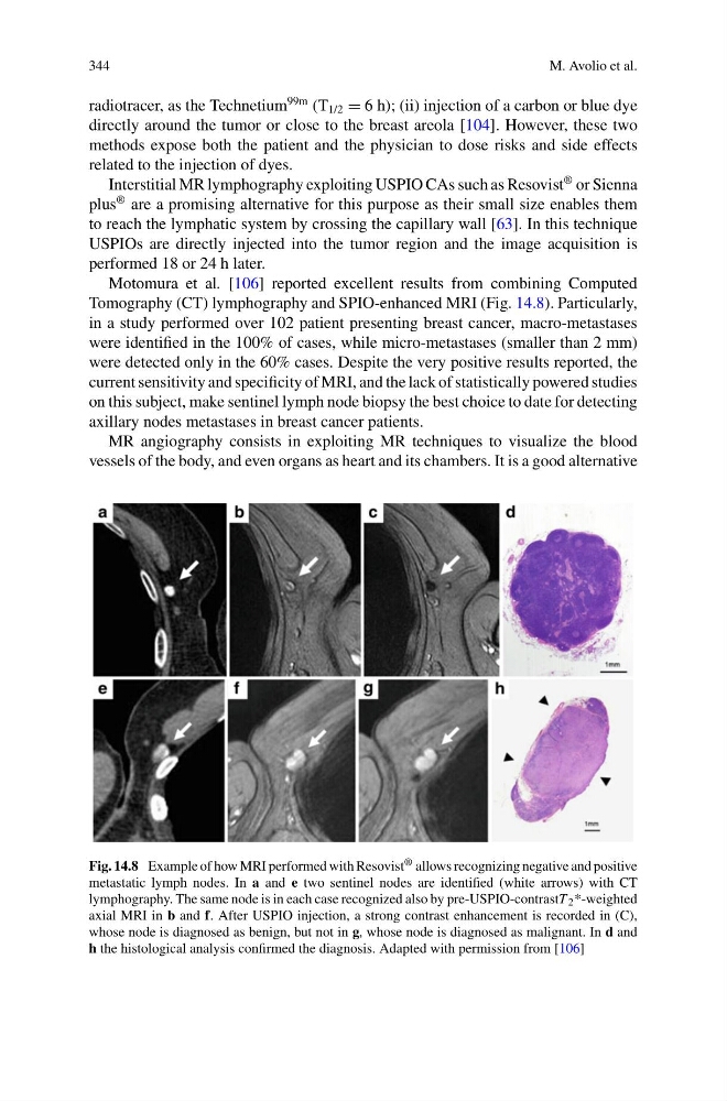
Motomura et al. [106] reported excellent results from combining Computed
Tomography (CT) lymphography and SPIO-enhanced MRI (Fig. 14.8). Particularly,
in a study performed over 102 patient presenting breast cancer, macro-metastases
were identified in the 100% of cases, while micro-metastases (smaller than 2 mm)
were detected only in the 60% cases. Despite the very positive results reported, the
current sensitivity and specificity of MRI, and the lack of statistically powered studies
on this subject, make sentinel lymph node biopsy the best choice to date for detecting
axillary nodes metastases in breast cancer patients.
MR angiography consists in exploiting MR techniques to visualize the blood
vessels of the body, and even organs as heart and its chambers. It is a good alternative
Fig. 14.8 Example of how MRI performed with Resovist ® allows recognizing negative and positive
metastatic lymph nodes. In a and e two sentinel nodes are identified (white arrows) with CT
lymphography. The same node is in each case recognized also by pre-USPIO-contrastT 2 *-weighted
axial MRI in b and f. After USPIO injection, a strong contrast enhancement is recorded in (C),
whose node is diagnosed as benign, but not in g, whose node is diagnosed as malignant. In d and
h the histological analysis confirmed the diagnosis. Adapted with permission from [106]
M. Avolio et al.
radiotracer, as the Technetium
99m (T 1/2 = 6 h); (ii) injection of a carbon or blue dye
directly around the tumor or close to the breast areola [104]. However, these two
methods expose both the patient and the physician to dose risks and side effects
related to the injection of dyes.
Interstitial MR lymphography exploiting USPIO CAs such as Resovist
® or Sienna
plus
® are a promising alternative for this purpose as their small size enables them
to reach the lymphatic system by crossing the capillary wall [63]. In this technique
USPIOs are directly injected into the tumor region and the image acquisition is
performed 18 or 24 h later.
Motomura et al. [106] reported excellent results from combining Computed
Tomography (CT) lymphography and SPIO-enhanced MRI (Fig. 14.8). Particularly,
in a study performed over 102 patient presenting breast cancer, macro-metastases
were identified in the 100% of cases, while micro-metastases (smaller than 2 mm)
were detected only in the 60% cases. Despite the very positive results reported, the
current sensitivity and specificity of MRI, and the lack of statistically powered studies
on this subject, make sentinel lymph node biopsy the best choice to date for detecting
axillary nodes metastases in breast cancer patients.
MR angiography consists in exploiting MR techniques to visualize the blood
vessels of the body, and even organs as heart and its chambers. It is a good alternative
Fig. 14.8 Example of how MRI performed with Resovist ® allows recognizing negative and positive
metastatic lymph nodes. In a and e two sentinel nodes are identified (white arrows) with CT
lymphography. The same node is in each case recognized also by pre-USPIO-contrastT 2 *-weighted
axial MRI in b and f. After USPIO injection, a strong contrast enhancement is recorded in (C),
whose node is diagnosed as benign, but not in g, whose node is diagnosed as malignant. In d and
h the histological analysis confirmed the diagnosis. Adapted with permission from [106]