

10 Measuring Atomic Magnetic Moments in Magnetic Nanostructures …
253
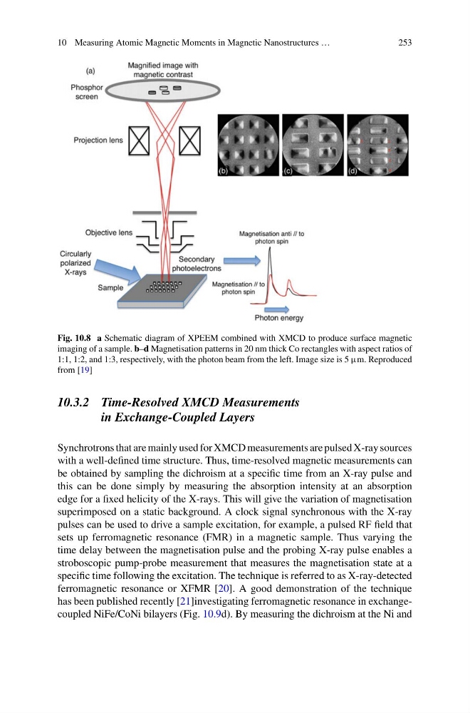
Fig. 10.8 a Schematic diagram of XPEEM combined with XMCD to produce surface magnetic
imaging of a sample. b–d Magnetisation patterns in 20 nm thick Co rectangles with aspect ratios of
1:1, 1:2, and 1:3, respectively, with the photon beam from the left. Image size is 5 μm. Reproduced
from [19]
10.3.2 Time-Resolved XMCD Measurements
in Exchange-Coupled Layers
Synchrotrons that are mainly used for XMCD measurements are pulsed X-ray sources
with a well-defined time structure. Thus, time-resolved magnetic measurements can
be obtained by sampling the dichroism at a specific time from an X-ray pulse and
this can be done simply by measuring the absorption intensity at an absorption
edge for a fixed helicity of the X-rays. This will give the variation of magnetisation
superimposed on a static background. A clock signal synchronous with the X-ray
pulses can be used to drive a sample excitation, for example, a pulsed RF field that
sets up ferromagnetic resonance (FMR) in a magnetic sample. Thus varying the
time delay between the magnetisation pulse and the probing X-ray pulse enables a
stroboscopic pump-probe measurement that measures the magnetisation state at a
specific time following the excitation. The technique is referred to as X-ray-detected
ferromagnetic resonance or XFMR [20]. A good demonstration of the technique
has been published recently [21]investigating ferromagnetic resonance in exchangecoupled NiFe/CoNi bilayers (Fig. 10.9d). By measuring the dichroism at the Ni and
253
Fig. 10.8 a Schematic diagram of XPEEM combined with XMCD to produce surface magnetic
imaging of a sample. b–d Magnetisation patterns in 20 nm thick Co rectangles with aspect ratios of
1:1, 1:2, and 1:3, respectively, with the photon beam from the left. Image size is 5 μm. Reproduced
from [19]
10.3.2 Time-Resolved XMCD Measurements
in Exchange-Coupled Layers
Synchrotrons that are mainly used for XMCD measurements are pulsed X-ray sources
with a well-defined time structure. Thus, time-resolved magnetic measurements can
be obtained by sampling the dichroism at a specific time from an X-ray pulse and
this can be done simply by measuring the absorption intensity at an absorption
edge for a fixed helicity of the X-rays. This will give the variation of magnetisation
superimposed on a static background. A clock signal synchronous with the X-ray
pulses can be used to drive a sample excitation, for example, a pulsed RF field that
sets up ferromagnetic resonance (FMR) in a magnetic sample. Thus varying the
time delay between the magnetisation pulse and the probing X-ray pulse enables a
stroboscopic pump-probe measurement that measures the magnetisation state at a
specific time following the excitation. The technique is referred to as X-ray-detected
ferromagnetic resonance or XFMR [20]. A good demonstration of the technique
has been published recently [21]investigating ferromagnetic resonance in exchangecoupled NiFe/CoNi bilayers (Fig. 10.9d). By measuring the dichroism at the Ni and