

230
A. Kleibert
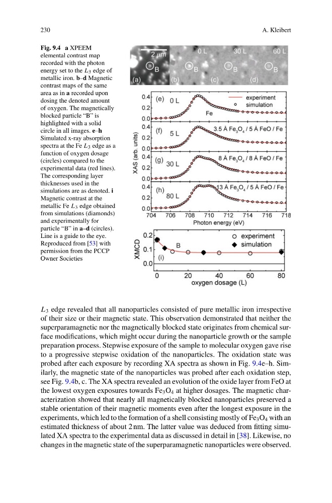
Fig. 9.4 a XPEEM
elemental contrast map
recorded with the photon
energy set to the L 3 edge of
metallic iron. b–d Magnetic
contrast maps of the same
area as in a recorded upon
dosing the denoted amount
of oxygen. The magnetically
blocked particle “B” is
highlighted with a solid
circle in all images. e–h
Simulated x-ray absorption
spectra at the Fe L 3 edge as a
function of oxygen dosage
(circles) compared to the
experimental data (red lines).
The corresponding layer
thicknesses used in the
simulations are as denoted. i
Magnetic contrast at the
metallic Fe L 3 edge obtained
from simulations (diamonds)
and experimentally for
particle “B” in a–d (circles).
Line is a guide to the eye.
Reproduced from [53] with
permission from the PCCP
Owner Societies
L 3 edge revealed that all nanoparticles consisted of pure metallic iron irrespective
of their size or their magnetic state. This observation demonstrated that neither the
superparamagnetic nor the magnetically blocked state originates from chemical surface modifications, which might occur during the nanoparticle growth or the sample
preparation process. Stepwise exposure of the sample to molecular oxygen gave rise
to a progressive stepwise oxidation of the nanoparticles. The oxidation state was
probed after each exposure by recording XA spectra as shown in Fig. 9.4e–h. Similarly, the magnetic state of the nanoparticles was probed after each oxidation step,
see Fig. 9.4b, c. The XA spectra revealed an evolution of the oxide layer from FeO at
the lowest oxygen exposures towards Fe 3 O 4 at higher dosages. The magnetic characterization showed that nearly all magnetically blocked nanoparticles preserved a
stable orientation of their magnetic moments even after the longest exposure in the
experiments, which led to the formation of a shell consisting mostly of Fe 3 O 4 with an
estimated thickness of about 2 nm. The latter value was deduced from fitting simulated XA spectra to the experimental data as discussed in detail in [38]. Likewise, no
changes in the magnetic state of the superparamagnetic nanoparticles were observed.
A. Kleibert
Fig. 9.4 a XPEEM
elemental contrast map
recorded with the photon
energy set to the L 3 edge of
metallic iron. b–d Magnetic
contrast maps of the same
area as in a recorded upon
dosing the denoted amount
of oxygen. The magnetically
blocked particle “B” is
highlighted with a solid
circle in all images. e–h
Simulated x-ray absorption
spectra at the Fe L 3 edge as a
function of oxygen dosage
(circles) compared to the
experimental data (red lines).
The corresponding layer
thicknesses used in the
simulations are as denoted. i
Magnetic contrast at the
metallic Fe L 3 edge obtained
from simulations (diamonds)
and experimentally for
particle “B” in a–d (circles).
Line is a guide to the eye.
Reproduced from [53] with
permission from the PCCP
Owner Societies
L 3 edge revealed that all nanoparticles consisted of pure metallic iron irrespective
of their size or their magnetic state. This observation demonstrated that neither the
superparamagnetic nor the magnetically blocked state originates from chemical surface modifications, which might occur during the nanoparticle growth or the sample
preparation process. Stepwise exposure of the sample to molecular oxygen gave rise
to a progressive stepwise oxidation of the nanoparticles. The oxidation state was
probed after each exposure by recording XA spectra as shown in Fig. 9.4e–h. Similarly, the magnetic state of the nanoparticles was probed after each oxidation step,
see Fig. 9.4b, c. The XA spectra revealed an evolution of the oxide layer from FeO at
the lowest oxygen exposures towards Fe 3 O 4 at higher dosages. The magnetic characterization showed that nearly all magnetically blocked nanoparticles preserved a
stable orientation of their magnetic moments even after the longest exposure in the
experiments, which led to the formation of a shell consisting mostly of Fe 3 O 4 with an
estimated thickness of about 2 nm. The latter value was deduced from fitting simulated XA spectra to the experimental data as discussed in detail in [38]. Likewise, no
changes in the magnetic state of the superparamagnetic nanoparticles were observed.