

9 Magnetism of Individual Nanoparticles Probed by X-Ray …
223
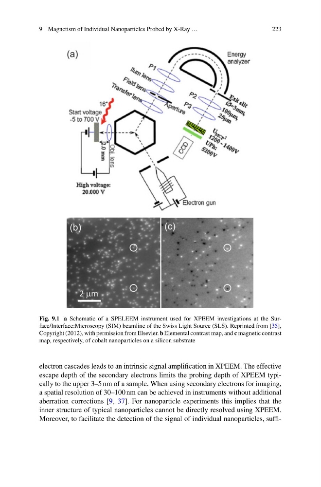
Fig. 9.1 a Schematic of a SPELEEM instrument used for XPEEM investigations at the Surface/Interface:Microscopy (SIM) beamline of the Swiss Light Source (SLS). Reprinted from [35],
Copyright (2012), with permission from Elsevier. b Elemental contrast map, and c magnetic contrast
map, respectively, of cobalt nanoparticles on a silicon substrate
electron cascades leads to an intrinsic signal amplification in XPEEM. The effective
escape depth of the secondary electrons limits the probing depth of XPEEM typically to the upper 3–5 nm of a sample. When using secondary electrons for imaging,
a spatial resolution of 30–100 nm can be achieved in instruments without additional
aberration corrections [9, 37]. For nanoparticle experiments this implies that the
inner structure of typical nanoparticles cannot be directly resolved using XPEEM.
Moreover, to facilitate the detection of the signal of individual nanoparticles, suffi-
223
Fig. 9.1 a Schematic of a SPELEEM instrument used for XPEEM investigations at the Surface/Interface:Microscopy (SIM) beamline of the Swiss Light Source (SLS). Reprinted from [35],
Copyright (2012), with permission from Elsevier. b Elemental contrast map, and c magnetic contrast
map, respectively, of cobalt nanoparticles on a silicon substrate
electron cascades leads to an intrinsic signal amplification in XPEEM. The effective
escape depth of the secondary electrons limits the probing depth of XPEEM typically to the upper 3–5 nm of a sample. When using secondary electrons for imaging,
a spatial resolution of 30–100 nm can be achieved in instruments without additional
aberration corrections [9, 37]. For nanoparticle experiments this implies that the
inner structure of typical nanoparticles cannot be directly resolved using XPEEM.
Moreover, to facilitate the detection of the signal of individual nanoparticles, suffi-