

14
M. A. Asson-Batres
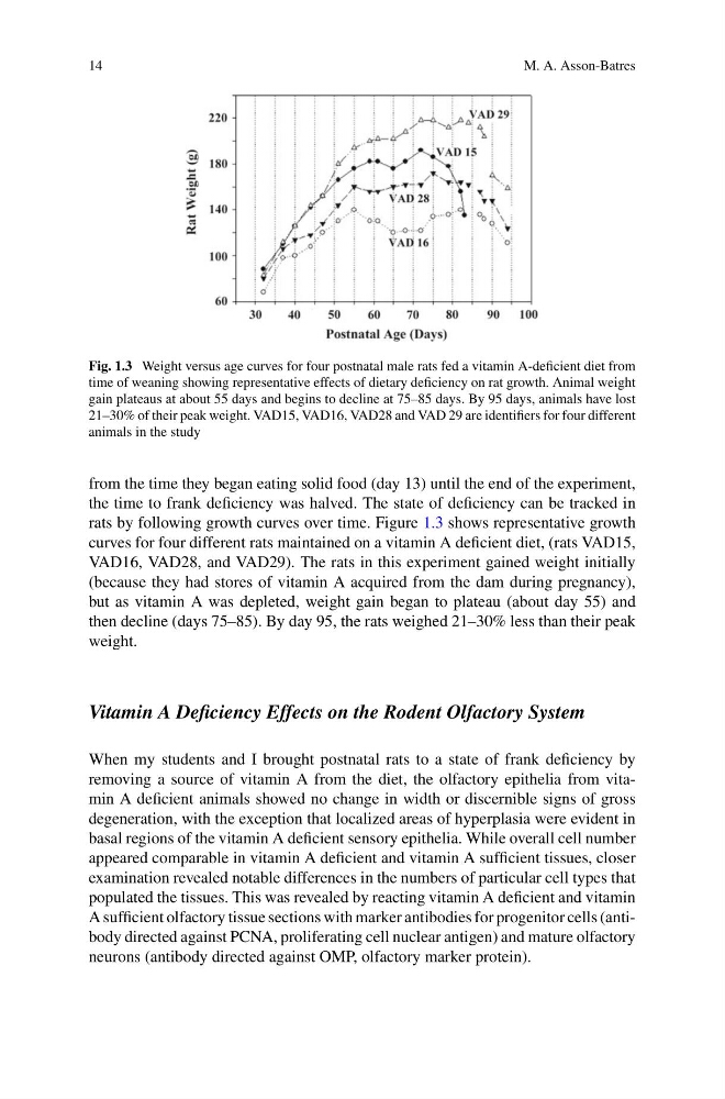
Fig. 1.3 Weight versus age curves for four postnatal male rats fed a vitamin A-deficient diet from
time of weaning showing representative effects of dietary deficiency on rat growth. Animal weight
gain plateaus at about 55 days and begins to decline at 75–85 days. By 95 days, animals have lost
21–30% of their peak weight. VAD15, VAD16, VAD28 and VAD 29 are identifiers for four different
animals in the study
from the time they began eating solid food (day 13) until the end of the experiment,
the time to frank deficiency was halved. The state of deficiency can be tracked in
rats by following growth curves over time. Figure 1.3 shows representative growth
curves for four different rats maintained on a vitamin A deficient diet, (rats VAD15,
VAD16, VAD28, and VAD29). The rats in this experiment gained weight initially
(because they had stores of vitamin A acquired from the dam during pregnancy),
but as vitamin A was depleted, weight gain began to plateau (about day 55) and
then decline (days 75–85). By day 95, the rats weighed 21–30% less than their peak
weight.
Vitamin A Deficiency Effects on the Rodent Olfactory System
When my students and I brought postnatal rats to a state of frank deficiency by
removing a source of vitamin A from the diet, the olfactory epithelia from vitamin A deficient animals showed no change in width or discernible signs of gross
degeneration, with the exception that localized areas of hyperplasia were evident in
basal regions of the vitamin A deficient sensory epithelia. While overall cell number
appeared comparable in vitamin A deficient and vitamin A sufficient tissues, closer
examination revealed notable differences in the numbers of particular cell types that
populated the tissues. This was revealed by reacting vitamin A deficient and vitamin
A sufficient olfactory tissue sections with marker antibodies for progenitor cells (antibody directed against PCNA, proliferating cell nuclear antigen) and mature olfactory
neurons (antibody directed against OMP, olfactory marker protein).
M. A. Asson-Batres
Fig. 1.3 Weight versus age curves for four postnatal male rats fed a vitamin A-deficient diet from
time of weaning showing representative effects of dietary deficiency on rat growth. Animal weight
gain plateaus at about 55 days and begins to decline at 75–85 days. By 95 days, animals have lost
21–30% of their peak weight. VAD15, VAD16, VAD28 and VAD 29 are identifiers for four different
animals in the study
from the time they began eating solid food (day 13) until the end of the experiment,
the time to frank deficiency was halved. The state of deficiency can be tracked in
rats by following growth curves over time. Figure 1.3 shows representative growth
curves for four different rats maintained on a vitamin A deficient diet, (rats VAD15,
VAD16, VAD28, and VAD29). The rats in this experiment gained weight initially
(because they had stores of vitamin A acquired from the dam during pregnancy),
but as vitamin A was depleted, weight gain began to plateau (about day 55) and
then decline (days 75–85). By day 95, the rats weighed 21–30% less than their peak
weight.
Vitamin A Deficiency Effects on the Rodent Olfactory System
When my students and I brought postnatal rats to a state of frank deficiency by
removing a source of vitamin A from the diet, the olfactory epithelia from vitamin A deficient animals showed no change in width or discernible signs of gross
degeneration, with the exception that localized areas of hyperplasia were evident in
basal regions of the vitamin A deficient sensory epithelia. While overall cell number
appeared comparable in vitamin A deficient and vitamin A sufficient tissues, closer
examination revealed notable differences in the numbers of particular cell types that
populated the tissues. This was revealed by reacting vitamin A deficient and vitamin
A sufficient olfactory tissue sections with marker antibodies for progenitor cells (antibody directed against PCNA, proliferating cell nuclear antigen) and mature olfactory
neurons (antibody directed against OMP, olfactory marker protein).