

178
W. R. Jackman and Y. Gibert
How important is RA signaling for controlling the most important aspects of tooth
development: where teeth form, how many teeth are present, and the size and shape
that they take? How much of the diversity of these three aspects of the dentition of
various species can be explained by evolutionary changes in RA cell signaling during
development? Specific studies in the zebrafish model system and related species have
begun to address these questions. To begin, a closer look at some of the early RA tooth
developmental studies will help to put these more recent findings into a historical
context.
History
Tooth Formation
To better understand specific aspects of tooth formation, it is necessary to consider
the primary tissue and cell types that are involved. The most highly conserved and
important aspects of teeth in vertebrates are dentin and enamel which form from
odontoblast and ameloblast cells respectively. Dentin is a bone-like tissue made
up of a semi-porous calcium phosphate-based extracellular matrix with a moderate
percentage of the extremely hard mineral hydroxyapatite (Peyer and Zangerl 1968).
Dentin comprises the bulk of most vertebrate teeth and is situated towards the center
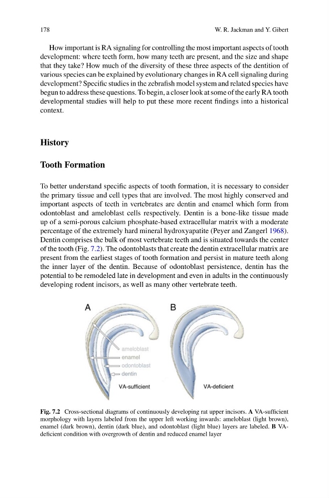
of the tooth (Fig. 7.2). The odontoblasts that create the dentin extracellular matrix are
present from the earliest stages of tooth formation and persist in mature teeth along
the inner layer of the dentin. Because of odontoblast persistence, dentin has the
potential to be remodeled late in development and even in adults in the continuously
developing rodent incisors, as well as many other vertebrate teeth.
Fig. 7.2 Cross-sectional diagrams of continuously developing rat upper incisors. A VA-sufficient
morphology with layers labeled from the upper left working inwards: ameloblast (light brown),
enamel (dark brown), dentin (dark blue), and odontoblast (light blue) layers are labeled. B VAdeficient condition with overgrowth of dentin and reduced enamel layer
W. R. Jackman and Y. Gibert
How important is RA signaling for controlling the most important aspects of tooth
development: where teeth form, how many teeth are present, and the size and shape
that they take? How much of the diversity of these three aspects of the dentition of
various species can be explained by evolutionary changes in RA cell signaling during
development? Specific studies in the zebrafish model system and related species have
begun to address these questions. To begin, a closer look at some of the early RA tooth
developmental studies will help to put these more recent findings into a historical
context.
History
Tooth Formation
To better understand specific aspects of tooth formation, it is necessary to consider
the primary tissue and cell types that are involved. The most highly conserved and
important aspects of teeth in vertebrates are dentin and enamel which form from
odontoblast and ameloblast cells respectively. Dentin is a bone-like tissue made
up of a semi-porous calcium phosphate-based extracellular matrix with a moderate
percentage of the extremely hard mineral hydroxyapatite (Peyer and Zangerl 1968).
Dentin comprises the bulk of most vertebrate teeth and is situated towards the center
of the tooth (Fig. 7.2). The odontoblasts that create the dentin extracellular matrix are
present from the earliest stages of tooth formation and persist in mature teeth along
the inner layer of the dentin. Because of odontoblast persistence, dentin has the
potential to be remodeled late in development and even in adults in the continuously
developing rodent incisors, as well as many other vertebrate teeth.
Fig. 7.2 Cross-sectional diagrams of continuously developing rat upper incisors. A VA-sufficient
morphology with layers labeled from the upper left working inwards: ameloblast (light brown),
enamel (dark brown), dentin (dark blue), and odontoblast (light blue) layers are labeled. B VAdeficient condition with overgrowth of dentin and reduced enamel layer