

132
E. Perl and J. S. Waxman
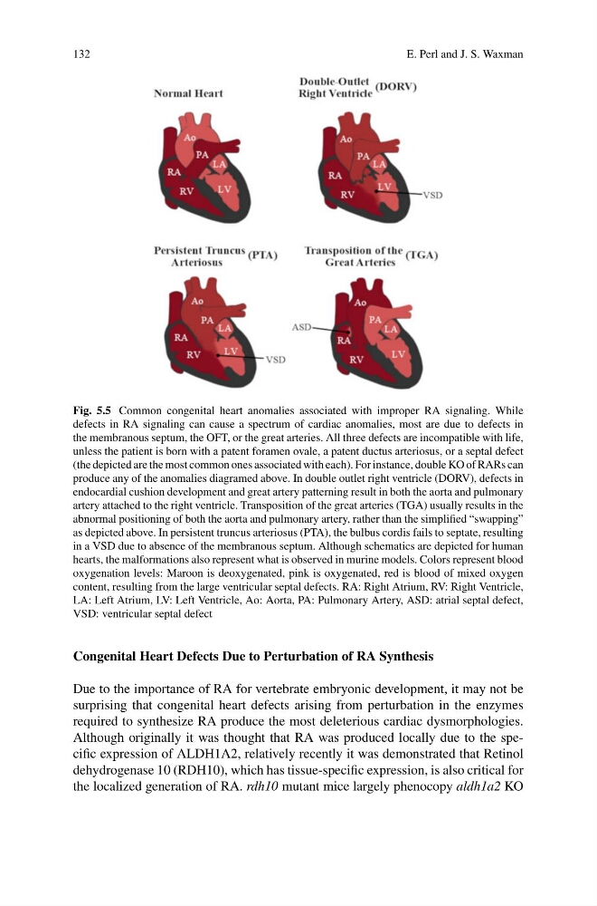
Fig. 5.5 Common congenital heart anomalies associated with improper RA signaling. While
defects in RA signaling can cause a spectrum of cardiac anomalies, most are due to defects in
the membranous septum, the OFT, or the great arteries. All three defects are incompatible with life,
unless the patient is born with a patent foramen ovale, a patent ductus arteriosus, or a septal defect
(the depicted are the most common ones associated with each). For instance, double KO of RARs can
produce any of the anomalies diagramed above. In double outlet right ventricle (DORV), defects in
endocardial cushion development and great artery patterning result in both the aorta and pulmonary
artery attached to the right ventricle. Transposition of the great arteries (TGA) usually results in the
abnormal positioning of both the aorta and pulmonary artery, rather than the simplified “swapping”
as depicted above. In persistent truncus arteriosus (PTA), the bulbus cordis fails to septate, resulting
in a VSD due to absence of the membranous septum. Although schematics are depicted for human
hearts, the malformations also represent what is observed in murine models. Colors represent blood
oxygenation levels: Maroon is deoxygenated, pink is oxygenated, red is blood of mixed oxygen
content, resulting from the large ventricular septal defects. RA: Right Atrium, RV: Right Ventricle,
LA: Left Atrium, LV: Left Ventricle, Ao: Aorta, PA: Pulmonary Artery, ASD: atrial septal defect,
VSD: ventricular septal defect
Congenital Heart Defects Due to Perturbation of RA Synthesis
Due to the importance of RA for vertebrate embryonic development, it may not be
surprising that congenital heart defects arising from perturbation in the enzymes
required to synthesize RA produce the most deleterious cardiac dysmorphologies.
Although originally it was thought that RA was produced locally due to the specific expression of ALDH1A2, relatively recently it was demonstrated that Retinol
dehydrogenase 10 (RDH10), which has tissue-specific expression, is also critical for
the localized generation of RA. rdh10 mutant mice largely phenocopy aldh1a2 KO
E. Perl and J. S. Waxman
Fig. 5.5 Common congenital heart anomalies associated with improper RA signaling. While
defects in RA signaling can cause a spectrum of cardiac anomalies, most are due to defects in
the membranous septum, the OFT, or the great arteries. All three defects are incompatible with life,
unless the patient is born with a patent foramen ovale, a patent ductus arteriosus, or a septal defect
(the depicted are the most common ones associated with each). For instance, double KO of RARs can
produce any of the anomalies diagramed above. In double outlet right ventricle (DORV), defects in
endocardial cushion development and great artery patterning result in both the aorta and pulmonary
artery attached to the right ventricle. Transposition of the great arteries (TGA) usually results in the
abnormal positioning of both the aorta and pulmonary artery, rather than the simplified “swapping”
as depicted above. In persistent truncus arteriosus (PTA), the bulbus cordis fails to septate, resulting
in a VSD due to absence of the membranous septum. Although schematics are depicted for human
hearts, the malformations also represent what is observed in murine models. Colors represent blood
oxygenation levels: Maroon is deoxygenated, pink is oxygenated, red is blood of mixed oxygen
content, resulting from the large ventricular septal defects. RA: Right Atrium, RV: Right Ventricle,
LA: Left Atrium, LV: Left Ventricle, Ao: Aorta, PA: Pulmonary Artery, ASD: atrial septal defect,
VSD: ventricular septal defect
Congenital Heart Defects Due to Perturbation of RA Synthesis
Due to the importance of RA for vertebrate embryonic development, it may not be
surprising that congenital heart defects arising from perturbation in the enzymes
required to synthesize RA produce the most deleterious cardiac dysmorphologies.
Although originally it was thought that RA was produced locally due to the specific expression of ALDH1A2, relatively recently it was demonstrated that Retinol
dehydrogenase 10 (RDH10), which has tissue-specific expression, is also critical for
the localized generation of RA. rdh10 mutant mice largely phenocopy aldh1a2 KO