

4 OPV with a Crystalline Organic Pigment Active Layer Up to 10 μm
7 9
With crystallization
Without crystallization
10-µm-thick crystalline OPV
( )
( )
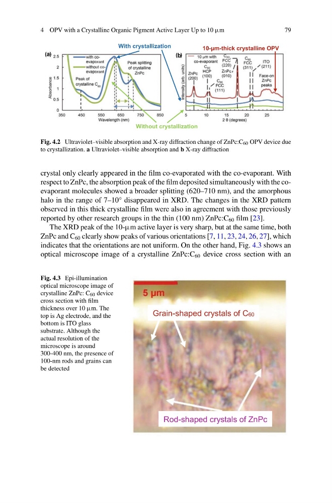
Fig. 4.2 Ultraviolet–visible absorption and X-ray diffraction change of ZnPc:C 60 OPV device due
to crystallization. a Ultraviolet–visible absorption and b X-ray diffraction
crystal only clearly appeared in the film co-evaporated with the co-evaporant. With
respect to ZnPc, the absorption peak of the film deposited simultaneously with the coevaporant molecules showed a broader splitting (620–710 nm), and the amorphous
halo in the range of 7–10° disappeared in XRD. The changes in the XRD pattern
observed in this thick crystalline film were also in agreement with those previously
reported by other research groups in the thin (100 nm) ZnPc:C 60 film [23].
The XRD peak of the 10-μm active layer is very sharp, but at the same time, both
ZnPc and C 60 clearly show peaks of various orientations [7, 11, 23, 24, 26, 27], which
indicates that the orientations are not uniform. On the other hand, Fig. 4.3 shows an
optical microscope image of a crystalline ZnPc:C 60 device cross section with an
Fig. 4.3 Epi-illumination
optical microscope image of
crystalline ZnPc: C 60 device
cross section with film
thickness over 10 μm. The
top is Ag electrode, and the
bottom is ITO glass
substrate. Although the
actual resolution of the
microscope is around
300-400 nm, the presence of
100-nm rods and grains can
be detected
Rod-shaped crystals of ZnPc
Grain-shaped crystals of C60
7 9
With crystallization
Without crystallization
10-µm-thick crystalline OPV
( )
( )
Fig. 4.2 Ultraviolet–visible absorption and X-ray diffraction change of ZnPc:C 60 OPV device due
to crystallization. a Ultraviolet–visible absorption and b X-ray diffraction
crystal only clearly appeared in the film co-evaporated with the co-evaporant. With
respect to ZnPc, the absorption peak of the film deposited simultaneously with the coevaporant molecules showed a broader splitting (620–710 nm), and the amorphous
halo in the range of 7–10° disappeared in XRD. The changes in the XRD pattern
observed in this thick crystalline film were also in agreement with those previously
reported by other research groups in the thin (100 nm) ZnPc:C 60 film [23].
The XRD peak of the 10-μm active layer is very sharp, but at the same time, both
ZnPc and C 60 clearly show peaks of various orientations [7, 11, 23, 24, 26, 27], which
indicates that the orientations are not uniform. On the other hand, Fig. 4.3 shows an
optical microscope image of a crystalline ZnPc:C 60 device cross section with an
Fig. 4.3 Epi-illumination
optical microscope image of
crystalline ZnPc: C 60 device
cross section with film
thickness over 10 μm. The
top is Ag electrode, and the
bottom is ITO glass
substrate. Although the
actual resolution of the
microscope is around
300-400 nm, the presence of
100-nm rods and grains can
be detected
Rod-shaped crystals of ZnPc
Grain-shaped crystals of C60