

A
SeCys158
His159
Arg407
E
B
C
W
Fe/S
Fe/S
Fe/S
Fe/S
D
W
Fe/S
Fe/S
Fe/S
Fe/S
F
G
SeCys192
His193
Arg441
SeCys192
His193
Arg441
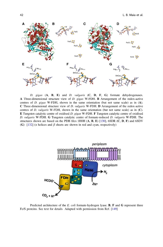
Fig. 6 D. gigas (A, B, E) and D. vulgaris (C, D, F, G) formate dehydrogenases.
A Three-dimensional structure view of D. gigas W-FDH. B Arrangement of the redox-active
centres of D. gigas W-FDH, shown in the same orientation (but not same scale) as in (A).
C Three-dimensional structure view of D. vulgaris W-FDH. D Arrangement of the redox-active
centres of D. vulgaris W-FDH, shown in the same orientation (but not same scale) as in (C).
E Tungsten catalytic centre of oxidised D. gigas W-FDH. F Tungsten catalytic centre of oxidised
D. vulgaris W-FDH. G Tungsten catalytic centre of formate-reduced D. vulgaris W-FDH. The
structures shown are based on the PDB files 1H0H (A, B, E) [130], 6SDR (C, D, F) and 6SDV
(G) [132] (a helices and b sheets are shown in red and cyan, respectively)
Fig. 7 Predicted architecture of the E. coli formate-hydrogen lyase. B, F and G represent three
Fe/S proteins. See text for details Adapted with permission from Ref. [149]
42
L. B. Maia et al.
SeCys158
His159
Arg407
E
B
C
W
Fe/S
Fe/S
Fe/S
Fe/S
D
W
Fe/S
Fe/S
Fe/S
Fe/S
F
G
SeCys192
His193
Arg441
SeCys192
His193
Arg441
Fig. 6 D. gigas (A, B, E) and D. vulgaris (C, D, F, G) formate dehydrogenases.
A Three-dimensional structure view of D. gigas W-FDH. B Arrangement of the redox-active
centres of D. gigas W-FDH, shown in the same orientation (but not same scale) as in (A).
C Three-dimensional structure view of D. vulgaris W-FDH. D Arrangement of the redox-active
centres of D. vulgaris W-FDH, shown in the same orientation (but not same scale) as in (C).
E Tungsten catalytic centre of oxidised D. gigas W-FDH. F Tungsten catalytic centre of oxidised
D. vulgaris W-FDH. G Tungsten catalytic centre of formate-reduced D. vulgaris W-FDH. The
structures shown are based on the PDB files 1H0H (A, B, E) [130], 6SDR (C, D, F) and 6SDV
(G) [132] (a helices and b sheets are shown in red and cyan, respectively)
Fig. 7 Predicted architecture of the E. coli formate-hydrogen lyase. B, F and G represent three
Fe/S proteins. See text for details Adapted with permission from Ref. [149]
42
L. B. Maia et al.