

for ligating and assembling metal nanocrystals. Colloidal dispersion of Co nanocrystals capped with fatty acids were found to self-assemble to yield hexagonally
ordered arrays similar to those obtained with alkanethiols [115, 116]. Similarly, Ag
nanocrystals capped with fatty acids of appropriate lengths yield cubic or hexagonal
close-packed structures [117, 118]. Schmid et al. [119] have reported an ordered
two-dimensional array of small Au 55 nanocrystals (diameter @ 1.4 nm) on a polymer film (see Figure 4.12). At the other end of the size-regime, large Au nanocrystals of 15–90 nm dimensions have also been organized into two-dimensional
arrays [120]. Arrays of AuaAg [62, 63] and FeaPt alloy nanocrystals [61] have been
obtained. Magic nuclearity Pd 561 nanocrystals, have been exploited to make PdaNi
core–shell particles with variable Ni loadings [121]. The nanocrystals so obtained
possess a core–shell structure, where a Ni layer covers a Pd seed. The magic nuclearity Pd 561 nanocrystals act as high quality seeds and promote the formation of
monodisperse PdaNi core–shell nanocrystals.
Arrays of Pd 561 Ni n (n upto 10,000 atoms) have been prepared after thiolizing the
core–shell nanocrystals as shown in Figure 4.13 [122]. By a simple extension of
this technique, arrays of triple layer nanocrystals of the form Pd 561 Ni 3000 Pd 1500
were also obtained. Methods to organize non-spherical metal nanocrystals into twodimensional arrays have met with very limited success. Thus, hexagonal Pt as well
as elongated silver nanocrystals have been organized into ordered two-dimensional
arrays [93, 123]. Interestingly, ordered two-dimensional lattices containing thiolized spherical Au particles of two different sizes have been reported by Kiely et al.
(see Figure 4.14) [113], who found that the nanocrystals of different radii follow the
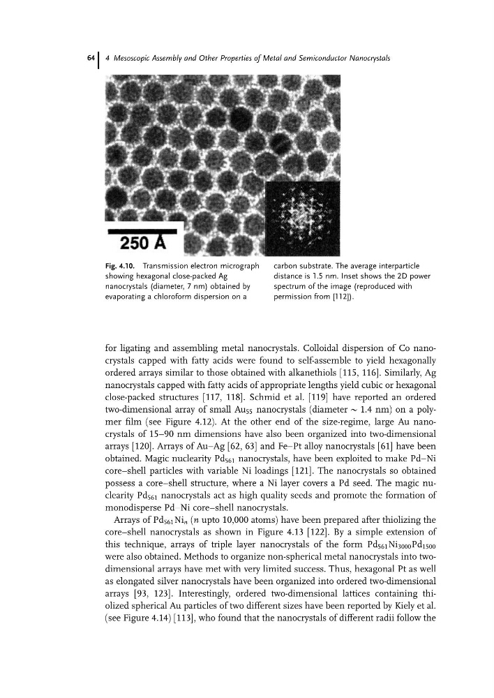
Fig. 4.10. Transmission electron micrograph
showing hexagonal close-packed Ag
nanocrystals (diameter, 7 nm) obtained by
evaporating a chloroform dispersion on a
carbon substrate. The average interparticle
distance is 1.5 nm. Inset shows the 2D power
spectrum of the image (reproduced with
permission from [112]).
4 Mesoscopic Assembly and Other Properties of Metal and Semiconductor Nanocrystals
64
ordered arrays similar to those obtained with alkanethiols [115, 116]. Similarly, Ag
nanocrystals capped with fatty acids of appropriate lengths yield cubic or hexagonal
close-packed structures [117, 118]. Schmid et al. [119] have reported an ordered
two-dimensional array of small Au 55 nanocrystals (diameter @ 1.4 nm) on a polymer film (see Figure 4.12). At the other end of the size-regime, large Au nanocrystals of 15–90 nm dimensions have also been organized into two-dimensional
arrays [120]. Arrays of AuaAg [62, 63] and FeaPt alloy nanocrystals [61] have been
obtained. Magic nuclearity Pd 561 nanocrystals, have been exploited to make PdaNi
core–shell particles with variable Ni loadings [121]. The nanocrystals so obtained
possess a core–shell structure, where a Ni layer covers a Pd seed. The magic nuclearity Pd 561 nanocrystals act as high quality seeds and promote the formation of
monodisperse PdaNi core–shell nanocrystals.
Arrays of Pd 561 Ni n (n upto 10,000 atoms) have been prepared after thiolizing the
core–shell nanocrystals as shown in Figure 4.13 [122]. By a simple extension of
this technique, arrays of triple layer nanocrystals of the form Pd 561 Ni 3000 Pd 1500
were also obtained. Methods to organize non-spherical metal nanocrystals into twodimensional arrays have met with very limited success. Thus, hexagonal Pt as well
as elongated silver nanocrystals have been organized into ordered two-dimensional
arrays [93, 123]. Interestingly, ordered two-dimensional lattices containing thiolized spherical Au particles of two different sizes have been reported by Kiely et al.
(see Figure 4.14) [113], who found that the nanocrystals of different radii follow the
Fig. 4.10. Transmission electron micrograph
showing hexagonal close-packed Ag
nanocrystals (diameter, 7 nm) obtained by
evaporating a chloroform dispersion on a
carbon substrate. The average interparticle
distance is 1.5 nm. Inset shows the 2D power
spectrum of the image (reproduced with
permission from [112]).
4 Mesoscopic Assembly and Other Properties of Metal and Semiconductor Nanocrystals
64