

It was suggested that the surface roughness plays a key role in facilitating charge
separation upon the substrate, which activates dispersed analytes for desorption/
ionization. A porous substructure, however, seems to function in some additional
capacity; the peak intensities of the mass spectrum from porous silicon measured
substantially greater that those from the other two surfaces. It was proposed that
diffusion from the porous substructure may serve to resupply the surface after a
laser pulse, increasing the absolute intensity of mass peaks and improving the
signal-to-noise ratio of the analysis. Desorption/ionization from porous silicon was
also accomplished by irradiation with an infrared (IR) laser source (IR-DIOS-MS)
(Figure 16.23), supporting a mechanism consistent with the heating-induced desorption of preformed surface ions. This mode of DIOS-MS succeeded only for
cooled specimens and apparently yielded finer results when water and acetic acid
vapors were deliberately leaked into the instrument chamber. The effects of low
temperature and vapors suggest that IR-DIOS-MS may proceed through a physisorbed solvent matrix that absorbs IR radiation and accommodates complete
transfer of energy to the dissolved analyte.
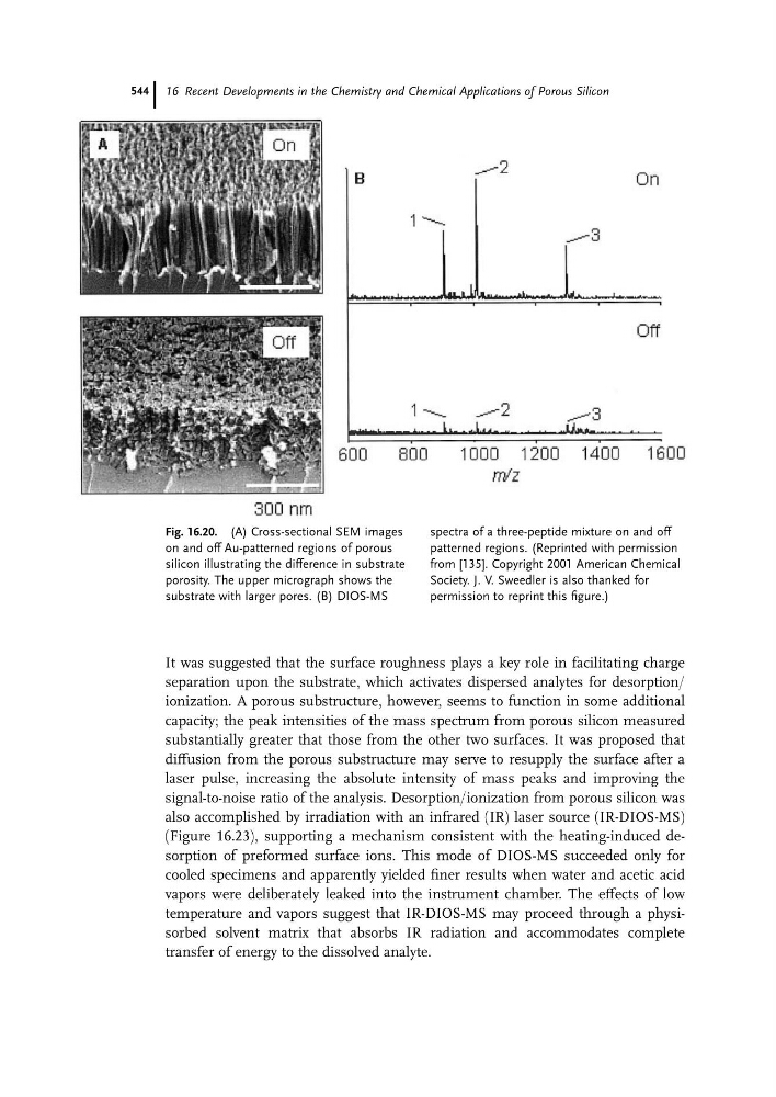
Fig. 16.20. (A) Cross-sectional SEM images
on and off Au-patterned regions of porous
silicon illustrating the difference in substrate
porosity. The upper micrograph shows the
substrate with larger pores. (B) DIOS-MS
spectra of a three-peptide mixture on and off
patterned regions. (Reprinted with permission
from [135]. Copyright 2001 American Chemical
Society. J. V. Sweedler is also thanked for
permission to reprint this figure.)
16 Recent Developments in the Chemistry and Chemical Applications of Porous Silicon
544