

rators to directly culture cells upon porous silicon and explore this material’s biocompatibility beyond the scope of inorganic biological materials [74–79]. Seeding
of porous silicon surfaces with Chinese hamster ovary cells was confirmed by
optical and rapid SEM after 24 h exposure. Despite their rapid degradation under
ambient conditions, these cells persistently remained attached to the substrates,
even after diligent rinsing, indicating that the cells adhered in some capacity to the
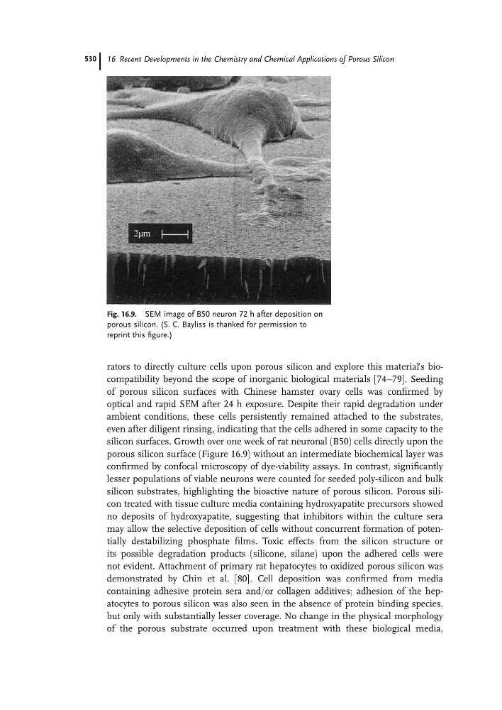
silicon surfaces. Growth over one week of rat neuronal (B50) cells directly upon the
porous silicon surface (Figure 16.9) without an intermediate biochemical layer was
confirmed by confocal microscopy of dye-viability assays. In contrast, significantly
lesser populations of viable neurons were counted for seeded poly-silicon and bulk
silicon substrates, highlighting the bioactive nature of porous silicon. Porous silicon treated with tissue culture media containing hydroxyapatite precursors showed
no deposits of hydroxyapatite, suggesting that inhibitors within the culture sera
may allow the selective deposition of cells without concurrent formation of potentially destabilizing phosphate films. Toxic effects from the silicon structure or
its possible degradation products (silicone, silane) upon the adhered cells were
not evident. Attachment of primary rat hepatocytes to oxidized porous silicon was
demonstrated by Chin et al. [80]. Cell deposition was confirmed from media
containing adhesive protein sera and/or collagen additives; adhesion of the hepatocytes to porous silicon was also seen in the absence of protein binding species,
but only with substantially lesser coverage. No change in the physical morphology
of the porous substrate occurred upon treatment with these biological media,
Fig. 16.9. SEM image of B50 neuron 72 h after deposition on
porous silicon. (S. C. Bayliss is thanked for permission to
reprint this figure.)
16 Recent Developments in the Chemistry and Chemical Applications of Porous Silicon
530