

by energy-dispersive spectroscopy (EDS), was observed to proceed almost exclusively upon the porous regions for brief exposures (30 min) to SBF under cathodic
conditions (Figure 16.8), although deposition across the entire surface during extended treatments (2 h) was also reported. Variation in the size, shape, and density
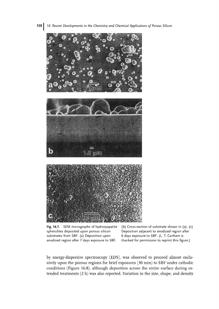
Fig. 16.7. SEM micrographs of hydroxyapatite
spherulites deposited upon porous silicon
substrates from SBF. (a) Deposition upon
anodized region after 7 days exposure to SBF.
(b) Cross-section of substrate shown in (a). (c)
Deposition adjacent to anodized region after
6 days exposure to SBF. (L. T. Canham is
thanked for permission to reprint this figure.)
16 Recent Developments in the Chemistry and Chemical Applications of Porous Silicon
528