

width of the Gaussian distribution of spacings between the particle centers was an
order of magnitude narrower in the concentrated dispersions compared to the dilute dispersions, consistent with more dense arrays in the concentrated dispersions
and a high degree of disorder in the dilute dispersions. No evidence for hexagonal
phases was found, under our conditions.
TEM images of air-dried dispersions prepared at low nanorod concentrations
(<1% by weight, including surfactant) showed mainly discrete nanoparticles
(Figure 9.7). Some short-range order involving side-on, end-to-side or end-to-end
aggregation was observed, presumably due to capillary forces associated with the
drying process [57]. At high nanorod concentrations (@5–10 wt%), in contrast,
microscopic smectic-like arrays of closely packed nanorods were observed (Figure
9.7). The arrays consisted of nanorods that were aligned parallel to each other in
micron-length rows, which in turn were stacked laterally to produce the higherorder superstructure. Such structures were observed predominantly at the edges of
dried droplets (apparent by visual inspection of the TEM grid as brown rings),
suggesting that capillary forces were responsible for the smectic-like organization [57]. In contrast, other areas of the TEM grid showed a predominance of
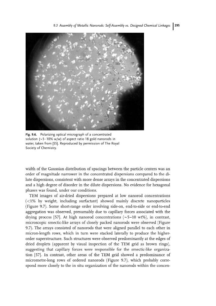
micrometre-long rows of ordered nanorods (Figure 9.7), which probably correspond more closely to the in situ organization of the nanorods within the concenFig. 9.6. Polarizing optical micrograph of a concentrated
solution (@5–10% w/w) of aspect ratio 18 gold nanorods in
water, taken from [55]. Reproduced by permission of The Royal
Society of Chemistry.
9.3 Assembly of Metallic Nanorods: Self-Assembly vs. Designed Chemical Linkages 295
order of magnitude narrower in the concentrated dispersions compared to the dilute dispersions, consistent with more dense arrays in the concentrated dispersions
and a high degree of disorder in the dilute dispersions. No evidence for hexagonal
phases was found, under our conditions.
TEM images of air-dried dispersions prepared at low nanorod concentrations
(<1% by weight, including surfactant) showed mainly discrete nanoparticles
(Figure 9.7). Some short-range order involving side-on, end-to-side or end-to-end
aggregation was observed, presumably due to capillary forces associated with the
drying process [57]. At high nanorod concentrations (@5–10 wt%), in contrast,
microscopic smectic-like arrays of closely packed nanorods were observed (Figure
9.7). The arrays consisted of nanorods that were aligned parallel to each other in
micron-length rows, which in turn were stacked laterally to produce the higherorder superstructure. Such structures were observed predominantly at the edges of
dried droplets (apparent by visual inspection of the TEM grid as brown rings),
suggesting that capillary forces were responsible for the smectic-like organization [57]. In contrast, other areas of the TEM grid showed a predominance of
micrometre-long rows of ordered nanorods (Figure 9.7), which probably correspond more closely to the in situ organization of the nanorods within the concenFig. 9.6. Polarizing optical micrograph of a concentrated
solution (@5–10% w/w) of aspect ratio 18 gold nanorods in
water, taken from [55]. Reproduced by permission of The Royal
Society of Chemistry.
9.3 Assembly of Metallic Nanorods: Self-Assembly vs. Designed Chemical Linkages 295