

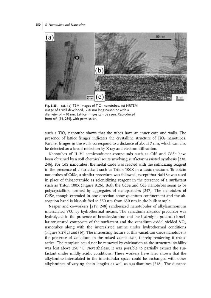
such a TiO 2 nanotube shows that the tubes have an inner core and walls. The
presence of lattice fringes indicates the crystalline structure of TiO 2 nanotubes.
Parallel fringes in the walls correspond to a distance of about 7 nm, which can also
be detected as a broad reflection by X-ray and electron diffraction.
Nanotubes of II–VI semiconductor compounds such as CdS and CdSe have
been obtained by a soft chemical route involving surfactant-assisted synthesis [238,
246]. For CdS nanotubes, the metal oxide was reacted with the sulfidizing reagent
in the presence of a surfactant such as Triton 100X in a basic medium. To obtain
nanotubes of CdSe, a similar procedure was followed, except that NaHSe was used
in place of thioacetamide as selenidizing reagent in the presence of a surfactant
such as Triton 100X (Figure 8.26). Both the CdSe and CdS nanotubes seem to be
polycrystalline, formed by aggregates of nanoparticles [247]. The nanotubes of
CdSe, though extended in one direction show quantum confinement and the absorption band is blue-shifted to 550 nm from 650 nm in the bulk sample.
Nesper and co-workers [219, 248] synthesized nanotubules of alkylammonium
intercalated VO x by hydrothermal means. The vanadium alkoxide precursor was
hydrolyzed in the presence of hexadecylamine and the hydrolysis product (lamellar structured composite of the surfactant and the vanadium oxide) yielded VO x
nanotubes along with the intercalated amine under hydrothermal conditions
(Figure 8.27(a) and (b)). The interesting feature of this vanadium oxide nanotube is
the presence of vanadium in the mixed valent state, thereby rendering it redoxactive. The template could not be removed by calcination as the structural stability
was lost above 250
C. Nevertheless, it was possible to partially extract the surfactant under mildly acidic conditions. These workers have later shown that the
alkylamine intercalated in the intertubular space could be exchanged with other
alkylamines of varying chain lengths as well as a,o-diamines [248]. The distance
Fig. 8.25. (a), (b) TEM images of TiO 2 nanotubes. (c) HRTEM
image of a well developed, @50 nm long nanotube with a
diameter of @10 nm. Lattice fringes can be seen. Reproduced
from ref. [24, 239], with permission.
8 Nanotubes and Nanowires
250
presence of lattice fringes indicates the crystalline structure of TiO 2 nanotubes.
Parallel fringes in the walls correspond to a distance of about 7 nm, which can also
be detected as a broad reflection by X-ray and electron diffraction.
Nanotubes of II–VI semiconductor compounds such as CdS and CdSe have
been obtained by a soft chemical route involving surfactant-assisted synthesis [238,
246]. For CdS nanotubes, the metal oxide was reacted with the sulfidizing reagent
in the presence of a surfactant such as Triton 100X in a basic medium. To obtain
nanotubes of CdSe, a similar procedure was followed, except that NaHSe was used
in place of thioacetamide as selenidizing reagent in the presence of a surfactant
such as Triton 100X (Figure 8.26). Both the CdSe and CdS nanotubes seem to be
polycrystalline, formed by aggregates of nanoparticles [247]. The nanotubes of
CdSe, though extended in one direction show quantum confinement and the absorption band is blue-shifted to 550 nm from 650 nm in the bulk sample.
Nesper and co-workers [219, 248] synthesized nanotubules of alkylammonium
intercalated VO x by hydrothermal means. The vanadium alkoxide precursor was
hydrolyzed in the presence of hexadecylamine and the hydrolysis product (lamellar structured composite of the surfactant and the vanadium oxide) yielded VO x
nanotubes along with the intercalated amine under hydrothermal conditions
(Figure 8.27(a) and (b)). The interesting feature of this vanadium oxide nanotube is
the presence of vanadium in the mixed valent state, thereby rendering it redoxactive. The template could not be removed by calcination as the structural stability
was lost above 250
C. Nevertheless, it was possible to partially extract the surfactant under mildly acidic conditions. These workers have later shown that the
alkylamine intercalated in the intertubular space could be exchanged with other
alkylamines of varying chain lengths as well as a,o-diamines [248]. The distance
Fig. 8.25. (a), (b) TEM images of TiO 2 nanotubes. (c) HRTEM
image of a well developed, @50 nm long nanotube with a
diameter of @10 nm. Lattice fringes can be seen. Reproduced
from ref. [24, 239], with permission.
8 Nanotubes and Nanowires
250