

transition metal is indeed found in the solid. The kinetics of the decomposition of
the carbonyl in the presence of the MSP was faster than in its absence (7 times
faster for Al-MCM-41). This is attributed to the large number of bubbles developed
due to the solid surface. The amount of MoO x in the solid Al-MCM-41 could reach
67 wt%. However, it was necessary to find the location of the MoO x particles. In
other words, to find out whether they have indeed entered the mesopores or can be
found outside the pores. This location was determined by mapping the TEM grid
employing TEM–EDAX measurements. The data clearly demonstrate [103] that
the Mo oxide phase is located inside the support’s pores and does not form separate particles up to an MoO 3 content of about 40–45 wt% upon ultrasonic deposition. High-resolution TEM (HRTEM) pictures [103] reveal that the MoO 3 deposition does not cause degradation of the Al-MCM-41 hexagonal pore structure. In
addition to the EDAX mapping technique, four other methods are used to support
these results. In the sample containing 67 wt% MoO x , large amounts of the excess
material are found outside the pores. Thus, a monolayer of MoO x particles is
strongly anchored to the walls, while the rest are found outside. Considering the
surface area of Al-MCM-41 used in the work and an Mo surface concentration of 5
Mo atoms nm
À2 , the geometrical closed, packed monolayer capacity corresponds to
50 wt% MoO 3 , which is in good agreement with our measurements. XPS measurements proved that chemical bonds are being formed between the silica and the
molybdena, forming SiaOaMo bonds.
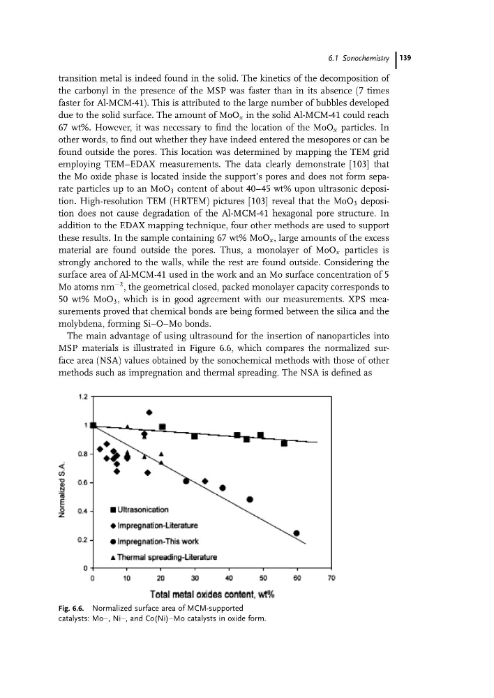
The main advantage of using ultrasound for the insertion of nanoparticles into
MSP materials is illustrated in Figure 6.6, which compares the normalized surface area (NSA) values obtained by the sonochemical methods with those of other
methods such as impregnation and thermal spreading. The NSA is defined as
Fig. 6.6. Normalized surface area of MCM-supported
catalysts: Moa, Nia, and Co(Ni)aMo catalysts in oxide form.
6.1 Sonochemistry 139
the carbonyl in the presence of the MSP was faster than in its absence (7 times
faster for Al-MCM-41). This is attributed to the large number of bubbles developed
due to the solid surface. The amount of MoO x in the solid Al-MCM-41 could reach
67 wt%. However, it was necessary to find the location of the MoO x particles. In
other words, to find out whether they have indeed entered the mesopores or can be
found outside the pores. This location was determined by mapping the TEM grid
employing TEM–EDAX measurements. The data clearly demonstrate [103] that
the Mo oxide phase is located inside the support’s pores and does not form separate particles up to an MoO 3 content of about 40–45 wt% upon ultrasonic deposition. High-resolution TEM (HRTEM) pictures [103] reveal that the MoO 3 deposition does not cause degradation of the Al-MCM-41 hexagonal pore structure. In
addition to the EDAX mapping technique, four other methods are used to support
these results. In the sample containing 67 wt% MoO x , large amounts of the excess
material are found outside the pores. Thus, a monolayer of MoO x particles is
strongly anchored to the walls, while the rest are found outside. Considering the
surface area of Al-MCM-41 used in the work and an Mo surface concentration of 5
Mo atoms nm
À2 , the geometrical closed, packed monolayer capacity corresponds to
50 wt% MoO 3 , which is in good agreement with our measurements. XPS measurements proved that chemical bonds are being formed between the silica and the
molybdena, forming SiaOaMo bonds.
The main advantage of using ultrasound for the insertion of nanoparticles into
MSP materials is illustrated in Figure 6.6, which compares the normalized surface area (NSA) values obtained by the sonochemical methods with those of other
methods such as impregnation and thermal spreading. The NSA is defined as
Fig. 6.6. Normalized surface area of MCM-supported
catalysts: Moa, Nia, and Co(Ni)aMo catalysts in oxide form.
6.1 Sonochemistry 139