

to form a kind of interface NiaOaAl bond. The connection positions further become nucleation centers for elemental nickel. After the sample is heated to high
temperatures, DRS and IR results showed the diffusion of nickel ions into the
vacant tetrahedral sites in alumina. At a higher temperature, an inversion process, the substitution of Ni
þ2 ions for part of the Al
þ3 at octahedral sites, takes
place, resulting finally in the formation of the disordered spinel phase. (4) Magnetization measurements show that the as-prepared sonication products are superparamagnetic due to the ultrafine nature of nickel particles.
In a few studies sonochemistry was used to coat polymers with nanosized particles [48–50]. Of these three reports one [50] dealt with metals, more specifically
with noble metals (Pt, Pd, and Au). In this research, metal colloids are adsorbed to
the surface of neutral functionalized polystyrene microspheres, PSMS. The authors report on the synthesis and characterization of catalytically important noble
monometallic colloids using various chemical and sonochemical methods. These
metal colloids are then adsorbed onto suitably functionalized PSMS. The metalimmobilized microspheres are reacted with a linker such as 4-mercaptobutyl
phosphonic acid and subsequently used to grow multilayers.
Lately, we have deposited silver nanoparticles with an average size of 5 nm on
the surface of preformed silica submicrospheres with the aid of power ultrasound
[51]. Ultrasound irradiation of a slurry of silica submicrospheres, silver nitrate, and
ammonia in an aqueous medium for 90 min under an atmosphere of argon to
hydrogen (95:5) yielded a silver–silica nanocomposite. By controlling the atmospheric and reaction conditions, we could achieve the deposition of metallic silver
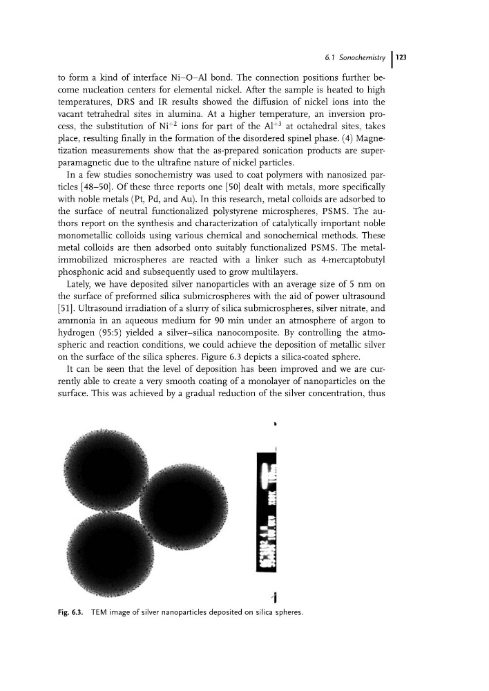
on the surface of the silica spheres. Figure 6.3 depicts a silica-coated sphere.
It can be seen that the level of deposition has been improved and we are currently able to create a very smooth coating of a monolayer of nanoparticles on the
surface. This was achieved by a gradual reduction of the silver concentration, thus
Fig. 6.3. TEM image of silver nanoparticles deposited on silica spheres.
6.1 Sonochemistry 123
temperatures, DRS and IR results showed the diffusion of nickel ions into the
vacant tetrahedral sites in alumina. At a higher temperature, an inversion process, the substitution of Ni
þ2 ions for part of the Al
þ3 at octahedral sites, takes
place, resulting finally in the formation of the disordered spinel phase. (4) Magnetization measurements show that the as-prepared sonication products are superparamagnetic due to the ultrafine nature of nickel particles.
In a few studies sonochemistry was used to coat polymers with nanosized particles [48–50]. Of these three reports one [50] dealt with metals, more specifically
with noble metals (Pt, Pd, and Au). In this research, metal colloids are adsorbed to
the surface of neutral functionalized polystyrene microspheres, PSMS. The authors report on the synthesis and characterization of catalytically important noble
monometallic colloids using various chemical and sonochemical methods. These
metal colloids are then adsorbed onto suitably functionalized PSMS. The metalimmobilized microspheres are reacted with a linker such as 4-mercaptobutyl
phosphonic acid and subsequently used to grow multilayers.
Lately, we have deposited silver nanoparticles with an average size of 5 nm on
the surface of preformed silica submicrospheres with the aid of power ultrasound
[51]. Ultrasound irradiation of a slurry of silica submicrospheres, silver nitrate, and
ammonia in an aqueous medium for 90 min under an atmosphere of argon to
hydrogen (95:5) yielded a silver–silica nanocomposite. By controlling the atmospheric and reaction conditions, we could achieve the deposition of metallic silver
on the surface of the silica spheres. Figure 6.3 depicts a silica-coated sphere.
It can be seen that the level of deposition has been improved and we are currently able to create a very smooth coating of a monolayer of nanoparticles on the
surface. This was achieved by a gradual reduction of the silver concentration, thus
Fig. 6.3. TEM image of silver nanoparticles deposited on silica spheres.
6.1 Sonochemistry 123