

2 Materials
Prepare all buffers and solutions using deionized water and analytical grade reagents. All cell culture steps are performed in a Biosafety
Level 2 laboratory. Manipulation of cells and cell culture reagents is
performed in a laminar flow hood using sterile consumables, unless
specified otherwise.
2.1 Cell Culture
1. HEK293T cells (European Collection of Animal Cell Culture,
Porton Down, UK).
2. Dulbecco’s Modified Eagle Medium with GlutaMAX™-I
(DMEM, 1Â). Add 50 mL fetal bovine serum (see Note 1)
and 5 mL nonessential amino acids 100Â to a 500 mL DMEM
bottle to obtain complete medium. Store at 4
C.
3. Cell detaching reagent: 0.05% trypsin, 0.05% EDTA in PBS.
4. Cell culture petri dishes (55 cm
2 ) or flasks (75 cm
2 ).
5. Falcon tubes.
6. Disposable serological pipettes.
7. CO 2 incubator, set at 37
C and 5% CO 2 .
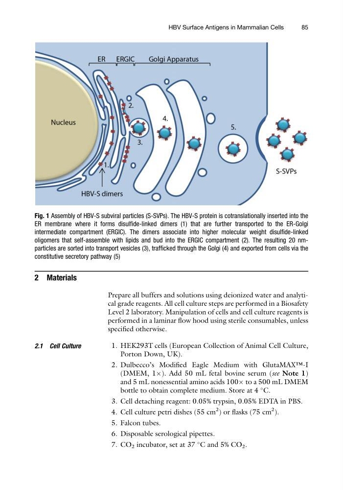
Fig. 1 Assembly of HBV-S subviral particles (S-SVPs). The HBV-S protein is cotranslationally inserted into the
ER membrane where it forms disulfide-linked dimers (1) that are further transported to the ER-Golgi
intermediate compartment (ERGIC). The dimers associate into higher molecular weight disulfide-linked
oligomers that self-assemble with lipids and bud into the ERGIC compartment (2). The resulting 20 nmparticles are sorted into transport vesicles (3), trafficked through the Golgi (4) and exported from cells via the
constitutive secretory pathway (5)
HBV Surface Antigens in Mammalian Cells
85
Prepare all buffers and solutions using deionized water and analytical grade reagents. All cell culture steps are performed in a Biosafety
Level 2 laboratory. Manipulation of cells and cell culture reagents is
performed in a laminar flow hood using sterile consumables, unless
specified otherwise.
2.1 Cell Culture
1. HEK293T cells (European Collection of Animal Cell Culture,
Porton Down, UK).
2. Dulbecco’s Modified Eagle Medium with GlutaMAX™-I
(DMEM, 1Â). Add 50 mL fetal bovine serum (see Note 1)
and 5 mL nonessential amino acids 100Â to a 500 mL DMEM
bottle to obtain complete medium. Store at 4
C.
3. Cell detaching reagent: 0.05% trypsin, 0.05% EDTA in PBS.
4. Cell culture petri dishes (55 cm
2 ) or flasks (75 cm
2 ).
5. Falcon tubes.
6. Disposable serological pipettes.
7. CO 2 incubator, set at 37
C and 5% CO 2 .
Fig. 1 Assembly of HBV-S subviral particles (S-SVPs). The HBV-S protein is cotranslationally inserted into the
ER membrane where it forms disulfide-linked dimers (1) that are further transported to the ER-Golgi
intermediate compartment (ERGIC). The dimers associate into higher molecular weight disulfide-linked
oligomers that self-assemble with lipids and bud into the ERGIC compartment (2). The resulting 20 nmparticles are sorted into transport vesicles (3), trafficked through the Golgi (4) and exported from cells via the
constitutive secretory pathway (5)
HBV Surface Antigens in Mammalian Cells
85