

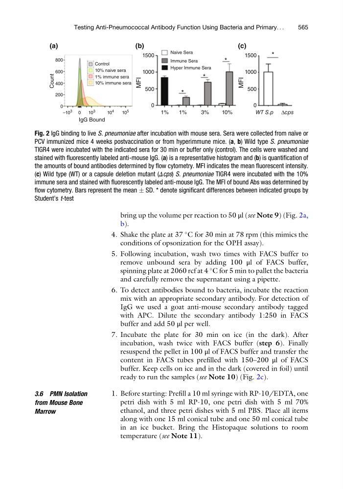
bring up the volume per reaction to 50 μl (see Note 9) (Fig. 2a,
b).
4. Shake the plate at 37
C for 30 min at 78 rpm (this mimics the
conditions of opsonization for the OPH assay).
5. Following incubation, wash two times with FACS buffer to
remove unbound sera by adding 100 μl of FACS buffer,
spinning plate at 2060 rcf at 4
C for 5 min to pallet the bacteria
and carefully remove the supernatant using a pipette.
6. To detect antibodies bound to bacteria, incubate the reaction
mix with an appropriate secondary antibody. For detection of
IgG we used a goat anti-mouse secondary antibody tagged
with APC. Dilute the secondary antibody 1:250 in FACS
buffer and add 50 μl per well.
7. Incubate the plate for 30 min on ice (in the dark). After
incubation, wash twice with FACS buffer (step 6). Finally
resuspend the pellet in 100 μl of FACS buffer and transfer the
content in FACS tubes prefilled with 150–200 μl of FACS
buffer. Keep cells on ice and in the dark (covered in foil) until
ready to run the samples (see Note 10) (Fig. 2c).
3.6 PMN Isolation
from Mouse Bone
Marrow
1. Before starting: Prefill a 10 ml syringe with RP-10/EDTA, one
petri dish with 5 ml RP-10, one petri dish with 5 ml 70%
ethanol, and three petri dishes with 5 ml PBS. Place all items
along with one 15 ml conical tube and one 50 ml conical tube
in an ice bucket. Bring the Histopaque solutions to room
temperature (see Note 11).
800
600
400
Control
(a)
(b)
(c)
Naive Sera
Immune Sera
Hyper Immune Sera
10% naive sera
1% immune sera
10% immune sera
1500
*
*
*
*
1000
500
MFI
0
1500
1000
500
MFI
0
1%
1%
3%
10%
WT S.p Δcps
200
0
–10
3
10
3
IgG Bound
Count
10
4
10
5
0
Fig. 2 IgG binding to live S. pneumoniae after incubation with mouse sera. Sera were collected from naı ¨ve or
PCV immunized mice 4 weeks postvaccination or from hyperimmune mice. (a, b) Wild type S. pneumoniae
TIGR4 were incubated with the indicated sera for 30 min or buffer only (control). The cells were washed and
stained with fluorescently labeled anti-mouse IgG. (a) is a representative histogram and (b) is quantification of
the amounts of bound antibodies determined by flow cytometry. MFI indicates the mean fluorescent intensity.
(c) Wild type (WT) or a capsule deletion mutant (Δcps) S. pneumoniae TIGR4 were incubated with the 10%
immune sera and stained with fluorescently labeled anti-mouse IgG. The MFI of bound Abs was determined by
flow cytometry. Bars represent the mean Æ SD. * denote significant differences between indicated groups by
Student’s t-test
Testing Anti-Pneumococcal Antibody Function Using Bacteria and Primary. . .
565
b).
4. Shake the plate at 37
C for 30 min at 78 rpm (this mimics the
conditions of opsonization for the OPH assay).
5. Following incubation, wash two times with FACS buffer to
remove unbound sera by adding 100 μl of FACS buffer,
spinning plate at 2060 rcf at 4
C for 5 min to pallet the bacteria
and carefully remove the supernatant using a pipette.
6. To detect antibodies bound to bacteria, incubate the reaction
mix with an appropriate secondary antibody. For detection of
IgG we used a goat anti-mouse secondary antibody tagged
with APC. Dilute the secondary antibody 1:250 in FACS
buffer and add 50 μl per well.
7. Incubate the plate for 30 min on ice (in the dark). After
incubation, wash twice with FACS buffer (step 6). Finally
resuspend the pellet in 100 μl of FACS buffer and transfer the
content in FACS tubes prefilled with 150–200 μl of FACS
buffer. Keep cells on ice and in the dark (covered in foil) until
ready to run the samples (see Note 10) (Fig. 2c).
3.6 PMN Isolation
from Mouse Bone
Marrow
1. Before starting: Prefill a 10 ml syringe with RP-10/EDTA, one
petri dish with 5 ml RP-10, one petri dish with 5 ml 70%
ethanol, and three petri dishes with 5 ml PBS. Place all items
along with one 15 ml conical tube and one 50 ml conical tube
in an ice bucket. Bring the Histopaque solutions to room
temperature (see Note 11).
800
600
400
Control
(a)
(b)
(c)
Naive Sera
Immune Sera
Hyper Immune Sera
10% naive sera
1% immune sera
10% immune sera
1500
*
*
*
*
1000
500
MFI
0
1500
1000
500
MFI
0
1%
1%
3%
10%
WT S.p Δcps
200
0
–10
3
10
3
IgG Bound
Count
10
4
10
5
0
Fig. 2 IgG binding to live S. pneumoniae after incubation with mouse sera. Sera were collected from naı ¨ve or
PCV immunized mice 4 weeks postvaccination or from hyperimmune mice. (a, b) Wild type S. pneumoniae
TIGR4 were incubated with the indicated sera for 30 min or buffer only (control). The cells were washed and
stained with fluorescently labeled anti-mouse IgG. (a) is a representative histogram and (b) is quantification of
the amounts of bound antibodies determined by flow cytometry. MFI indicates the mean fluorescent intensity.
(c) Wild type (WT) or a capsule deletion mutant (Δcps) S. pneumoniae TIGR4 were incubated with the 10%
immune sera and stained with fluorescently labeled anti-mouse IgG. The MFI of bound Abs was determined by
flow cytometry. Bars represent the mean Æ SD. * denote significant differences between indicated groups by
Student’s t-test
Testing Anti-Pneumococcal Antibody Function Using Bacteria and Primary. . .
565