

7. Add secondary anti-mouse IgG-Fc, IgG1, and IgG2a antibodies diluted 1:10,000 in PBS-T (100 μl/well), and incubate at
RT for 1 h.
8. Repeat step 2.
9. Bound antibody is detected by using p-nitrophenylphosphate
phosphatase as substrate.
10. Absorbances are measured at 405 nm using the PowereWave
XS2 plate reader with GEN5 software (BioTek, Winooski, VT).
3.2.3 Influenza Virus
Microneutralization Assay
1. Mouse sera are heat-inactivated at 56
for 30 min (see Note 3).
2. Dilute the mouse sera in serial dilutions.
3. Add 50 μl of each dilution and place in the wells of a microtiter
plate and mix with 50 μl (2 Â 10
4 PFU) of H1N1 influenza
virus A/PR/8/34 (PR8, from VR-95; ATCC, Manassas, VA).
4. Incubate the plate for 1 h at 37
, and 50 μl of the suspension
should be transferred onto Madin–Darby canine kidney
(MDCK) cells.
5. After 1 h of incubation at 37
in 5% CO 2 , the suspension
containing virus and antibody is removed, and 1% agarose (see
Note 4) in medium (minimum essential medium, 0.2% BSA,
1 μg/ml TPCK-treated trypsin) is added to each well.
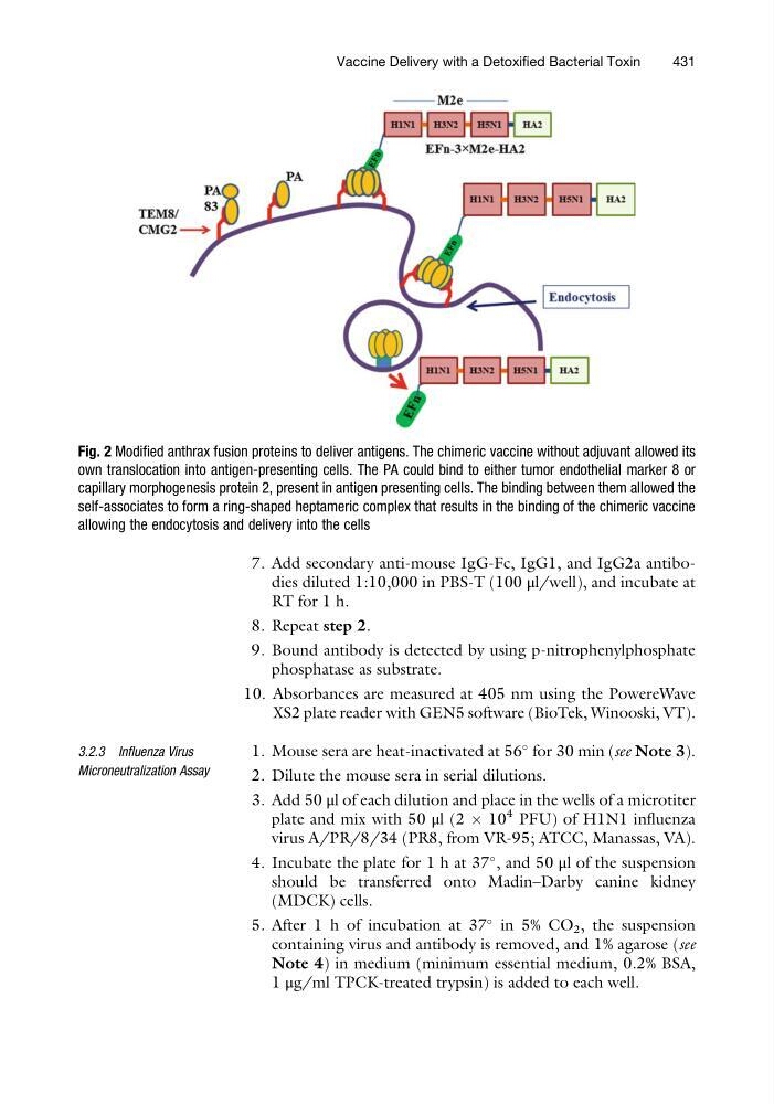
Fig. 2 Modified anthrax fusion proteins to deliver antigens. The chimeric vaccine without adjuvant allowed its
own translocation into antigen-presenting cells. The PA could bind to either tumor endothelial marker 8 or
capillary morphogenesis protein 2, present in antigen presenting cells. The binding between them allowed the
self-associates to form a ring-shaped heptameric complex that results in the binding of the chimeric vaccine
allowing the endocytosis and delivery into the cells
Vaccine Delivery with a Detoxified Bacterial Toxin
431
RT for 1 h.
8. Repeat step 2.
9. Bound antibody is detected by using p-nitrophenylphosphate
phosphatase as substrate.
10. Absorbances are measured at 405 nm using the PowereWave
XS2 plate reader with GEN5 software (BioTek, Winooski, VT).
3.2.3 Influenza Virus
Microneutralization Assay
1. Mouse sera are heat-inactivated at 56
for 30 min (see Note 3).
2. Dilute the mouse sera in serial dilutions.
3. Add 50 μl of each dilution and place in the wells of a microtiter
plate and mix with 50 μl (2 Â 10
4 PFU) of H1N1 influenza
virus A/PR/8/34 (PR8, from VR-95; ATCC, Manassas, VA).
4. Incubate the plate for 1 h at 37
, and 50 μl of the suspension
should be transferred onto Madin–Darby canine kidney
(MDCK) cells.
5. After 1 h of incubation at 37
in 5% CO 2 , the suspension
containing virus and antibody is removed, and 1% agarose (see
Note 4) in medium (minimum essential medium, 0.2% BSA,
1 μg/ml TPCK-treated trypsin) is added to each well.
Fig. 2 Modified anthrax fusion proteins to deliver antigens. The chimeric vaccine without adjuvant allowed its
own translocation into antigen-presenting cells. The PA could bind to either tumor endothelial marker 8 or
capillary morphogenesis protein 2, present in antigen presenting cells. The binding between them allowed the
self-associates to form a ring-shaped heptameric complex that results in the binding of the chimeric vaccine
allowing the endocytosis and delivery into the cells
Vaccine Delivery with a Detoxified Bacterial Toxin
431