

2. Purified EB samples are commonly quantified in inclusion
forming units (IFU) by in vitro assays [21]. However this
method does not count the many noninfectious EBs that will
still be stained by this protocol and will bind to and label cells.
Therefore it is advised to quantify the EBs prior to this protocol
using a method to measure absolute particle number or total
protein.
3. When pelleting EBs during the staining procedure, a microcentrifuge with a maximum speed of >20,000 Â g should be
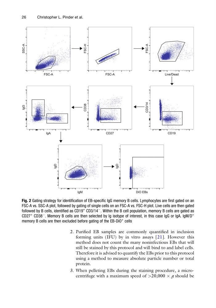
Fig. 2 Gating strategy for identification of EB-specific IgG memory B cells. Lymphocytes are first gated on an
FSC-A vs. SSC-A plot, followed by gating of single cells on an FSC-A vs. FSC-H plot. Live cells are then gated
followed by B cells, identified as CD19
+ CD3/14
À . Within the B cell population, memory B cells are gated as
CD27
+ CD38
À . Memory B cells are then selected by Ig isotype of interest, in this case IgG or IgA. IgM/D
+
memory B cells are then excluded before gating of the EB-DiO
+ cells
26
Christopher L. Pinder et al.
forming units (IFU) by in vitro assays [21]. However this
method does not count the many noninfectious EBs that will
still be stained by this protocol and will bind to and label cells.
Therefore it is advised to quantify the EBs prior to this protocol
using a method to measure absolute particle number or total
protein.
3. When pelleting EBs during the staining procedure, a microcentrifuge with a maximum speed of >20,000 Â g should be
Fig. 2 Gating strategy for identification of EB-specific IgG memory B cells. Lymphocytes are first gated on an
FSC-A vs. SSC-A plot, followed by gating of single cells on an FSC-A vs. FSC-H plot. Live cells are then gated
followed by B cells, identified as CD19
+ CD3/14
À . Within the B cell population, memory B cells are gated as
CD27
+ CD38
À . Memory B cells are then selected by Ig isotype of interest, in this case IgG or IgA. IgM/D
+
memory B cells are then excluded before gating of the EB-DiO
+ cells
26
Christopher L. Pinder et al.