

4. Preparation of mobile phase 2: 20 mM Tris–HCl (pH 8.0),
1 M NaCl. Transfer 20 mL of the 1 M Tris–HCl buffer and
200 mL of the 5 M NaCl solution to a 1 L volumetric flask and
add water to the graduation mark. Filter the buffer and transfer
it to a 1 L flask for autoclavation (see Note 8).
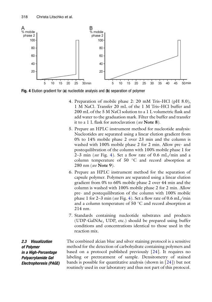
5. Prepare an HPLC instrument method for nucleotide analysis:
Nucleotides are separated using a linear elution gradient from
0% to 14% mobile phase 2 over 23 min and the column is
washed with 100% mobile phase 2 for 2 min. Allow pre- and
postequilibration of the column with 100% mobile phase 1 for
2–3 min (see Fig. 4). Set a flow rate of 0.6 mL/min and a
column temperature of 50
C and record absorption at
280 nm (see Note 9).
6. Prepare an HPLC instrument method for the separation of
capsule polymer. Polymers are separated using a linear elution
gradient from 0% to 60% mobile phase 2 over 44 min and the
column is washed with 100% mobile phase 2 for 2 min. Allow
pre- and postequilibration of the column with 100% mobile
phase 1 for 2–3 min (see Fig. 4). Set a flow rate of 0.6 mL/min
and a column temperature of 50
C and record absorption at
214 nm.
7. Standards containing nucleotide substrates and products
(UDP-GalNAc, UDP, etc.) should be prepared using buffer
conditions and concentrations identical to those used in the
reaction mix.
2.3 Visualization
of Polymer
in a High-Percentage
Polyacrylamide Gel
Electrophoresis (PAGE)
The combined alcian blue and silver staining protocol is a sensitive
method for the detection of carbohydrate containing polymers and
based on a protocol published previously [24]. It requires no
labeling or pretreatment of sample. Densitometry of stained
bands is possible for quantitative analysis (shown in [24]) but not
routinely used in our laboratory and thus not part of this protocol.
Fig. 4 Elution gradient for (a) nucleotide analysis and (b) separation of polymer
318
Christa Litschko et al.
1 M NaCl. Transfer 20 mL of the 1 M Tris–HCl buffer and
200 mL of the 5 M NaCl solution to a 1 L volumetric flask and
add water to the graduation mark. Filter the buffer and transfer
it to a 1 L flask for autoclavation (see Note 8).
5. Prepare an HPLC instrument method for nucleotide analysis:
Nucleotides are separated using a linear elution gradient from
0% to 14% mobile phase 2 over 23 min and the column is
washed with 100% mobile phase 2 for 2 min. Allow pre- and
postequilibration of the column with 100% mobile phase 1 for
2–3 min (see Fig. 4). Set a flow rate of 0.6 mL/min and a
column temperature of 50
C and record absorption at
280 nm (see Note 9).
6. Prepare an HPLC instrument method for the separation of
capsule polymer. Polymers are separated using a linear elution
gradient from 0% to 60% mobile phase 2 over 44 min and the
column is washed with 100% mobile phase 2 for 2 min. Allow
pre- and postequilibration of the column with 100% mobile
phase 1 for 2–3 min (see Fig. 4). Set a flow rate of 0.6 mL/min
and a column temperature of 50
C and record absorption at
214 nm.
7. Standards containing nucleotide substrates and products
(UDP-GalNAc, UDP, etc.) should be prepared using buffer
conditions and concentrations identical to those used in the
reaction mix.
2.3 Visualization
of Polymer
in a High-Percentage
Polyacrylamide Gel
Electrophoresis (PAGE)
The combined alcian blue and silver staining protocol is a sensitive
method for the detection of carbohydrate containing polymers and
based on a protocol published previously [24]. It requires no
labeling or pretreatment of sample. Densitometry of stained
bands is possible for quantitative analysis (shown in [24]) but not
routinely used in our laboratory and thus not part of this protocol.
Fig. 4 Elution gradient for (a) nucleotide analysis and (b) separation of polymer
318
Christa Litschko et al.