

3. Gently add 100 ng cloned transfer vector plasmid DNA to the
MAX Efficiency
® DH10-Bac™.
4. Place the vial with the bacteria suspension on ice for 30 min.
5. Induce a heat-shock at 42
C for 45 s without shaking to
enforce a plasmid uptake.
6. Incubate the bacteria on ice for an additional 2 min.
7. Add 750 μL S.O.C. Medium.
8. Incubate the bacteria at 37
C and 450 rpm for 4 h.
9. Add LB medium to dilute the suspension 1:10.
10. Seed 100 μL of the diluted bacteria suspension on the agar
plates containing kanamycin, tetracycline, X-Gal, and IPTG.
11. Incubate the plates overnight at 37
C.
12. Select white E. coli colonies and restreak them on fresh agar
plates containing kanamycin, tetracycline, X-Gal, and IPTG.
13. Incubate the agar plate overnight at 37
C.
3.2.2 Analysis
of the Recombinant Bacmid
Using LongAmp Taq
Polymerase
1. Pick one white colony from restreaked agar plates and suspend
it into tubes containing 5 mL LB medium and kanamycin,
tetracycline each—follow the procedure 6–10-times (meaning
that you have 6–10 colonies in culture at the end).
2. Incubate the liquid cultures at 37
C and 250 rpm overnight.
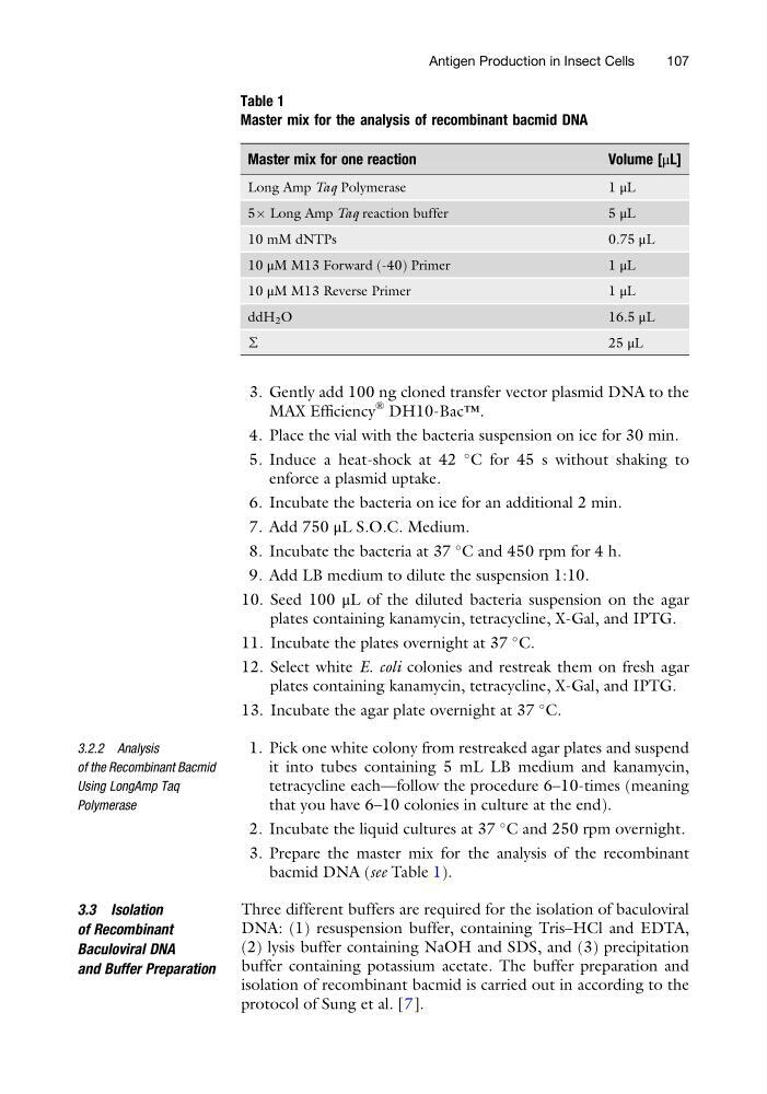
3. Prepare the master mix for the analysis of the recombinant
bacmid DNA (see Table 1).
3.3 Isolation
of Recombinant
Baculoviral DNA
and Buffer Preparation
Three different buffers are required for the isolation of baculoviral
DNA: (1) resuspension buffer, containing Tris–HCl and EDTA,
(2) lysis buffer containing NaOH and SDS, and (3) precipitation
buffer containing potassium acetate. The buffer preparation and
isolation of recombinant bacmid is carried out in according to the
protocol of Sung et al. [7].
Table 1
Master mix for the analysis of recombinant bacmid DNA
Master mix for one reaction
Volume [μL]
Long Amp Taq Polymerase
1 μL
5Â Long Amp Taq reaction buffer
5 μL
10 mM dNTPs
0.75 μL
10 μM M13 Forward (-40) Primer
1 μL
10 μM M13 Reverse Primer
1 μL
ddH 2 O
16.5 μL
∑
25 μL
Antigen Production in Insect Cells
107
MAX Efficiency
® DH10-Bac™.
4. Place the vial with the bacteria suspension on ice for 30 min.
5. Induce a heat-shock at 42
C for 45 s without shaking to
enforce a plasmid uptake.
6. Incubate the bacteria on ice for an additional 2 min.
7. Add 750 μL S.O.C. Medium.
8. Incubate the bacteria at 37
C and 450 rpm for 4 h.
9. Add LB medium to dilute the suspension 1:10.
10. Seed 100 μL of the diluted bacteria suspension on the agar
plates containing kanamycin, tetracycline, X-Gal, and IPTG.
11. Incubate the plates overnight at 37
C.
12. Select white E. coli colonies and restreak them on fresh agar
plates containing kanamycin, tetracycline, X-Gal, and IPTG.
13. Incubate the agar plate overnight at 37
C.
3.2.2 Analysis
of the Recombinant Bacmid
Using LongAmp Taq
Polymerase
1. Pick one white colony from restreaked agar plates and suspend
it into tubes containing 5 mL LB medium and kanamycin,
tetracycline each—follow the procedure 6–10-times (meaning
that you have 6–10 colonies in culture at the end).
2. Incubate the liquid cultures at 37
C and 250 rpm overnight.
3. Prepare the master mix for the analysis of the recombinant
bacmid DNA (see Table 1).
3.3 Isolation
of Recombinant
Baculoviral DNA
and Buffer Preparation
Three different buffers are required for the isolation of baculoviral
DNA: (1) resuspension buffer, containing Tris–HCl and EDTA,
(2) lysis buffer containing NaOH and SDS, and (3) precipitation
buffer containing potassium acetate. The buffer preparation and
isolation of recombinant bacmid is carried out in according to the
protocol of Sung et al. [7].
Table 1
Master mix for the analysis of recombinant bacmid DNA
Master mix for one reaction
Volume [μL]
Long Amp Taq Polymerase
1 μL
5Â Long Amp Taq reaction buffer
5 μL
10 mM dNTPs
0.75 μL
10 μM M13 Forward (-40) Primer
1 μL
10 μM M13 Reverse Primer
1 μL
ddH 2 O
16.5 μL
∑
25 μL
Antigen Production in Insect Cells
107