

11 Fluorescent Chemosensors
379
should be kept in mind that any molecule exhibiting high reactivity or affinity to the
analyte (even the dye itself) may act as a binding site. For example, cationic and/or
planar fluorophores tend to bind to DNA despite having no specific binding sites.
Thus, various molecular structural characteristics, such as the polarity, rigidity, and
hydrophobicity/hydrophilicity may direct the fluorophore to the analyte.
11.2.2 Representative Structures
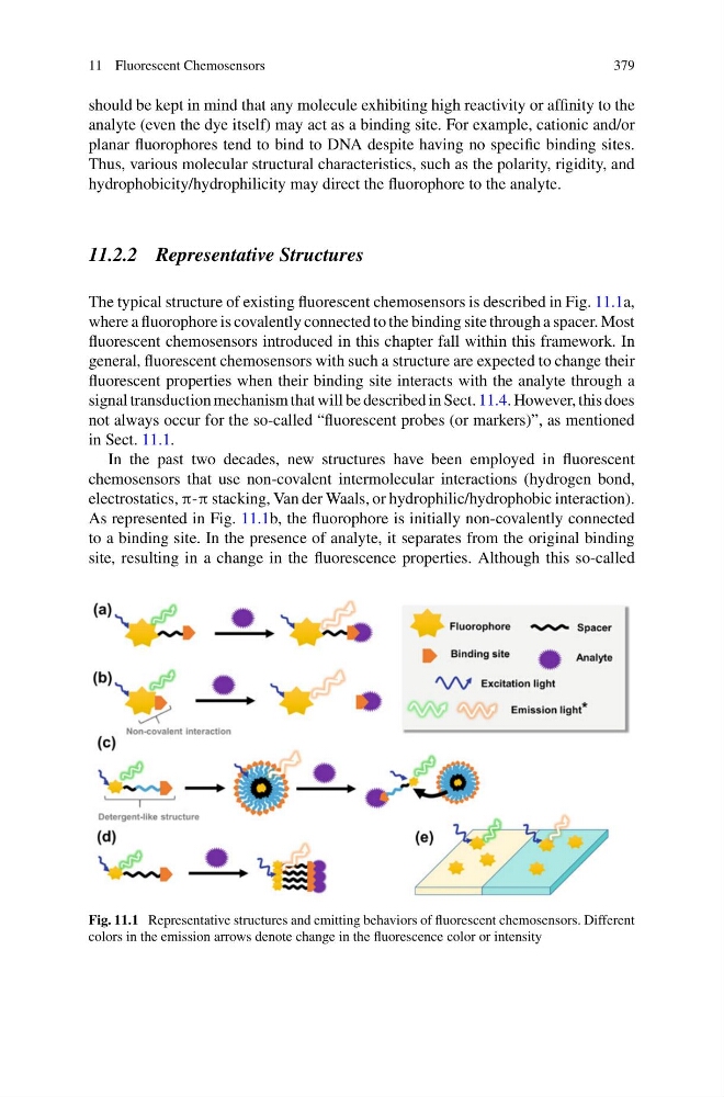
The typical structure of existing fluorescent chemosensors is described in Fig. 11.1a,
where a fluorophore is covalently connected to the binding site through a spacer. Most
fluorescent chemosensors introduced in this chapter fall within this framework. In
general, fluorescent chemosensors with such a structure are expected to change their
fluorescent properties when their binding site interacts with the analyte through a
signal transduction mechanism that will be described in Sect. 11.4. However, this does
not always occur for the so-called “fluorescent probes (or markers)”, as mentioned
in Sect. 11.1.
In the past two decades, new structures have been employed in fluorescent
chemosensors that use non-covalent intermolecular interactions (hydrogen bond,
electrostatics, π-π stacking, Van der Waals, or hydrophilic/hydrophobic interaction).
As represented in Fig. 11.1b, the fluorophore is initially non-covalently connected
to a binding site. In the presence of analyte, it separates from the original binding
site, resulting in a change in the fluorescence properties. Although this so-called
Fig. 11.1 Representative structures and emitting behaviors of fluorescent chemosensors. Different
colors in the emission arrows denote change in the fluorescence color or intensity
379
should be kept in mind that any molecule exhibiting high reactivity or affinity to the
analyte (even the dye itself) may act as a binding site. For example, cationic and/or
planar fluorophores tend to bind to DNA despite having no specific binding sites.
Thus, various molecular structural characteristics, such as the polarity, rigidity, and
hydrophobicity/hydrophilicity may direct the fluorophore to the analyte.
11.2.2 Representative Structures
The typical structure of existing fluorescent chemosensors is described in Fig. 11.1a,
where a fluorophore is covalently connected to the binding site through a spacer. Most
fluorescent chemosensors introduced in this chapter fall within this framework. In
general, fluorescent chemosensors with such a structure are expected to change their
fluorescent properties when their binding site interacts with the analyte through a
signal transduction mechanism that will be described in Sect. 11.4. However, this does
not always occur for the so-called “fluorescent probes (or markers)”, as mentioned
in Sect. 11.1.
In the past two decades, new structures have been employed in fluorescent
chemosensors that use non-covalent intermolecular interactions (hydrogen bond,
electrostatics, π-π stacking, Van der Waals, or hydrophilic/hydrophobic interaction).
As represented in Fig. 11.1b, the fluorophore is initially non-covalently connected
to a binding site. In the presence of analyte, it separates from the original binding
site, resulting in a change in the fluorescence properties. Although this so-called
Fig. 11.1 Representative structures and emitting behaviors of fluorescent chemosensors. Different
colors in the emission arrows denote change in the fluorescence color or intensity