

6 Molecular and Crystal Structures of Polymorphic …
235
(a) α I
(b) β
(c) γ
(d) α II
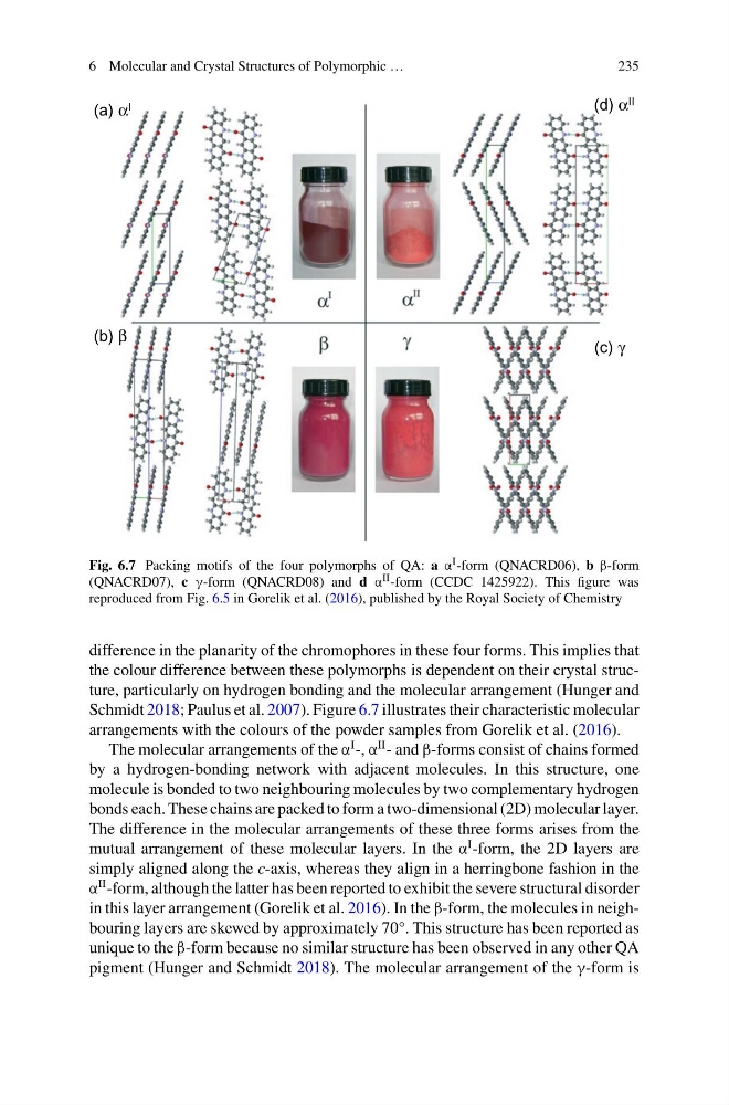
Fig. 6.7 Packing motifs of the four polymorphs of QA: a α I -form (QNACRD06), b β-form
(QNACRD07), c γ-form (QNACRD08) and d α II -form (CCDC 1425922). This figure was
reproduced from Fig. 6.5 in Gorelik et al. (2016), published by the Royal Society of Chemistry
difference in the planarity of the chromophores in these four forms. This implies that
the colour difference between these polymorphs is dependent on their crystal structure, particularly on hydrogen bonding and the molecular arrangement (Hunger and
Schmidt 2018; Paulus et al. 2007). Figure 6.7 illustrates their characteristic molecular
arrangements with the colours of the powder samples from Gorelik et al. (2016).
The molecular arrangements of the α
I -, α
II - and β-forms consist of chains formed
by a hydrogen-bonding network with adjacent molecules. In this structure, one
molecule is bonded to two neighbouring molecules by two complementary hydrogen
bonds each. These chains are packed to form a two-dimensional (2D) molecular layer.
The difference in the molecular arrangements of these three forms arises from the
mutual arrangement of these molecular layers. In the α
I -form, the 2D layers are
simply aligned along the c-axis, whereas they align in a herringbone fashion in the
α
II -form, although the latter has been reported to exhibit the severe structural disorder
in this layer arrangement (Gorelik et al. 2016). In the β-form, the molecules in neighbouring layers are skewed by approximately 70°. This structure has been reported as
unique to the β-form because no similar structure has been observed in any other QA
pigment (Hunger and Schmidt 2018). The molecular arrangement of the γ-form is
235
(a) α I
(b) β
(c) γ
(d) α II
Fig. 6.7 Packing motifs of the four polymorphs of QA: a α I -form (QNACRD06), b β-form
(QNACRD07), c γ-form (QNACRD08) and d α II -form (CCDC 1425922). This figure was
reproduced from Fig. 6.5 in Gorelik et al. (2016), published by the Royal Society of Chemistry
difference in the planarity of the chromophores in these four forms. This implies that
the colour difference between these polymorphs is dependent on their crystal structure, particularly on hydrogen bonding and the molecular arrangement (Hunger and
Schmidt 2018; Paulus et al. 2007). Figure 6.7 illustrates their characteristic molecular
arrangements with the colours of the powder samples from Gorelik et al. (2016).
The molecular arrangements of the α
I -, α
II - and β-forms consist of chains formed
by a hydrogen-bonding network with adjacent molecules. In this structure, one
molecule is bonded to two neighbouring molecules by two complementary hydrogen
bonds each. These chains are packed to form a two-dimensional (2D) molecular layer.
The difference in the molecular arrangements of these three forms arises from the
mutual arrangement of these molecular layers. In the α
I -form, the 2D layers are
simply aligned along the c-axis, whereas they align in a herringbone fashion in the
α
II -form, although the latter has been reported to exhibit the severe structural disorder
in this layer arrangement (Gorelik et al. 2016). In the β-form, the molecules in neighbouring layers are skewed by approximately 70°. This structure has been reported as
unique to the β-form because no similar structure has been observed in any other QA
pigment (Hunger and Schmidt 2018). The molecular arrangement of the γ-form is