

222 ◾ Fundamental Food Microbiology
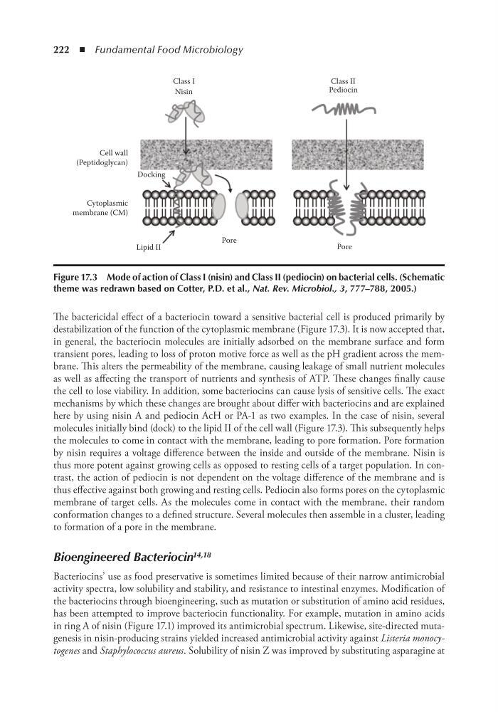
The bactericidal effect of a bacteriocin toward a sensitive bacterial cell is produced primarily by
destabilization of the function of the cytoplasmic membrane (Figure 17.3). It is now accepted that,
in general, the bacteriocin molecules are initially adsorbed on the membrane surface and form
transient pores, leading to loss of proton motive force as well as the pH gradient across the membrane. This alters the permeability of the membrane, causing leakage of small nutrient molecules
as well as affecting the transport of nutrients and synthesis of ATP. These changes finally cause
the cell to lose viability. In addition, some bacteriocins can cause lysis of sensitive cells. The exact
mechanisms by which these changes are brought about differ with bacteriocins and are explained
here by using nisin A and pediocin AcH or PA-1 as two examples. In the case of nisin, several
molecules initially bind (dock) to the lipid II of the cell wall (Figure 17.3). This subsequently helps
the molecules to come in contact with the membrane, leading to pore formation. Pore formation
by nisin requires a voltage difference between the inside and outside of the membrane. Nisin is
thus more potent against growing cells as opposed to resting cells of a target population. In contrast, the action of pediocin is not dependent on the voltage difference of the membrane and is
thus effective against both growing and resting cells. Pediocin also forms pores on the cytoplasmic
membrane of target cells. As the molecules come in contact with the membrane, their random
conformation changes to a defined structure. Several molecules then assemble in a cluster, leading
to formation of a pore in the membrane.
Bioengineered Bacteriocin 14,18
Bacteriocins’ use as food preservative is sometimes limited because of their narrow antimicrobial
activity spectra, low solubility and stability, and resistance to intestinal enzymes. Modification of
the bacteriocins through bioengineering, such as mutation or substitution of amino acid residues,
has been attempted to improve bacteriocin functionality. For example, mutation in amino acids
in ring A of nisin (Figure 17.1) improved its antimicrobial spectrum. Likewise, site-directed mutagenesis in nisin-producing strains yielded increased antimicrobial activity against Listeria monocytogenes and Staphylococcus aureus. Solubility of nisin Z was improved by substituting asparagine at
Class I
Class II
Nisin
Pediocin
Cell wall
(Peptidoglycan)
Docking
Cytoplasmic
membrane (CM)
Lipid II
Pore
Pore
Figure 17.3 Mode of action of Class i (nisin) and Class ii (pediocin) on bacterial cells. (Schematic
theme was redrawn based on Cotter, P.D. et al., Nat. Rev. Microbiol., 3, 777–788, 2005.)
The bactericidal effect of a bacteriocin toward a sensitive bacterial cell is produced primarily by
destabilization of the function of the cytoplasmic membrane (Figure 17.3). It is now accepted that,
in general, the bacteriocin molecules are initially adsorbed on the membrane surface and form
transient pores, leading to loss of proton motive force as well as the pH gradient across the membrane. This alters the permeability of the membrane, causing leakage of small nutrient molecules
as well as affecting the transport of nutrients and synthesis of ATP. These changes finally cause
the cell to lose viability. In addition, some bacteriocins can cause lysis of sensitive cells. The exact
mechanisms by which these changes are brought about differ with bacteriocins and are explained
here by using nisin A and pediocin AcH or PA-1 as two examples. In the case of nisin, several
molecules initially bind (dock) to the lipid II of the cell wall (Figure 17.3). This subsequently helps
the molecules to come in contact with the membrane, leading to pore formation. Pore formation
by nisin requires a voltage difference between the inside and outside of the membrane. Nisin is
thus more potent against growing cells as opposed to resting cells of a target population. In contrast, the action of pediocin is not dependent on the voltage difference of the membrane and is
thus effective against both growing and resting cells. Pediocin also forms pores on the cytoplasmic
membrane of target cells. As the molecules come in contact with the membrane, their random
conformation changes to a defined structure. Several molecules then assemble in a cluster, leading
to formation of a pore in the membrane.
Bioengineered Bacteriocin 14,18
Bacteriocins’ use as food preservative is sometimes limited because of their narrow antimicrobial
activity spectra, low solubility and stability, and resistance to intestinal enzymes. Modification of
the bacteriocins through bioengineering, such as mutation or substitution of amino acid residues,
has been attempted to improve bacteriocin functionality. For example, mutation in amino acids
in ring A of nisin (Figure 17.1) improved its antimicrobial spectrum. Likewise, site-directed mutagenesis in nisin-producing strains yielded increased antimicrobial activity against Listeria monocytogenes and Staphylococcus aureus. Solubility of nisin Z was improved by substituting asparagine at
Class I
Class II
Nisin
Pediocin
Cell wall
(Peptidoglycan)
Docking
Cytoplasmic
membrane (CM)
Lipid II
Pore
Pore
Figure 17.3 Mode of action of Class i (nisin) and Class ii (pediocin) on bacterial cells. (Schematic
theme was redrawn based on Cotter, P.D. et al., Nat. Rev. Microbiol., 3, 777–788, 2005.)