

313
trapping substrates or other molecules that bind to the enzyme to become part of the
structure, which is fundamental for structure-function studies (Rhodes 2000).
Atomic’ resolution at ≥1.2°A resolution allows the placement of atoms with
fewer geometrical restraints and gives a better picture of the protein structure.
Advances in X-ray sources and cryo-crystallography have led to increasing numbers of structures solved at these high resolutions (Kleywegt et al. 1996). The threedimensional representation of the protein may be displayed in a molecular structure
viewer as a model that was created by the crystallographer to be chemically realistic
and to match the observed electron density as precisely as possible. The resolution
of a crystal structure is measured in angstrom and refers to the minimum distance
between two points that can be distinguished. Although there is a large number of
quality assessment methods available, resolution is a straightforward and robust
parameter to assess the quality of a protein structure model (Kleywegt et al. 2004).
Nuclear Magnetic Resonance (NMR) Spectroscopy
NMR spectroscopy is a powerful tool for elucidating the structure–function relationships of substrates, peptides, proteins and in particular enzymes. It yields
detailed information regarding structure of enzyme and the specific ligands which
bind to the enzyme. The structure of ligands at the binding sites of enzymes and the
structure of enzyme–ligand complexes can also be obtained, as well as the dynamics
of the ligand and the associated structure of the protein binding site. The tertiary
structures of proteins can now be obtained independently of diffraction data in solution by homo nuclear and hetero nuclear multi-dimensional NMR. In principle one
can investigate the magnetic nuclei of each of the atoms within the molecule of the
enzyme (
1
H,
13
C,
15
N, …) or ligands which bind to the enzyme (
1
H,
19
F,
31
P,
13
C, …),
or of the environment of the active-site (solvent
1
H
2
O,
2
D
2
O,
23
Na,
39
K,
35
Cl, …)
(Monasterio 2014). Until recently, NMR spectroscopy has yielded structures of protein complexes with small and medium size (~30 to 40 kDa). Major breakthroughs
during recent past especially in isotope-labeling techniques, have enabled NMR
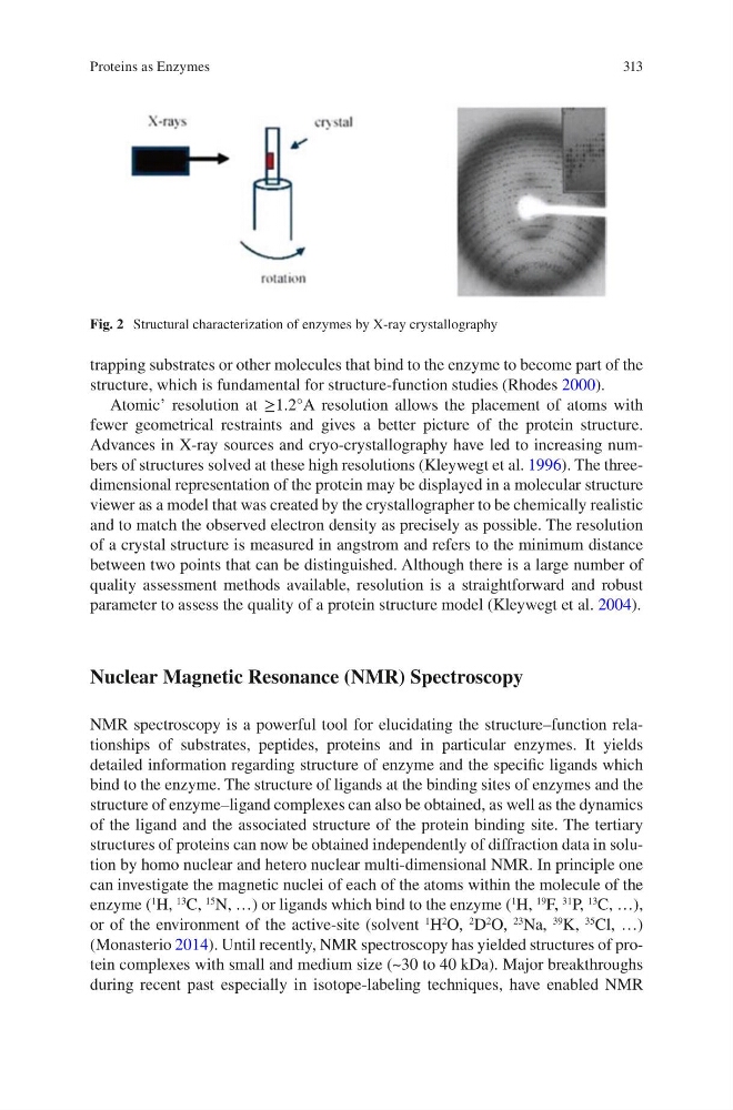
Fig. 2 Structural characterization of enzymes by X-ray crystallography
Proteins as Enzymes
trapping substrates or other molecules that bind to the enzyme to become part of the
structure, which is fundamental for structure-function studies (Rhodes 2000).
Atomic’ resolution at ≥1.2°A resolution allows the placement of atoms with
fewer geometrical restraints and gives a better picture of the protein structure.
Advances in X-ray sources and cryo-crystallography have led to increasing numbers of structures solved at these high resolutions (Kleywegt et al. 1996). The threedimensional representation of the protein may be displayed in a molecular structure
viewer as a model that was created by the crystallographer to be chemically realistic
and to match the observed electron density as precisely as possible. The resolution
of a crystal structure is measured in angstrom and refers to the minimum distance
between two points that can be distinguished. Although there is a large number of
quality assessment methods available, resolution is a straightforward and robust
parameter to assess the quality of a protein structure model (Kleywegt et al. 2004).
Nuclear Magnetic Resonance (NMR) Spectroscopy
NMR spectroscopy is a powerful tool for elucidating the structure–function relationships of substrates, peptides, proteins and in particular enzymes. It yields
detailed information regarding structure of enzyme and the specific ligands which
bind to the enzyme. The structure of ligands at the binding sites of enzymes and the
structure of enzyme–ligand complexes can also be obtained, as well as the dynamics
of the ligand and the associated structure of the protein binding site. The tertiary
structures of proteins can now be obtained independently of diffraction data in solution by homo nuclear and hetero nuclear multi-dimensional NMR. In principle one
can investigate the magnetic nuclei of each of the atoms within the molecule of the
enzyme (
1
H,
13
C,
15
N, …) or ligands which bind to the enzyme (
1
H,
19
F,
31
P,
13
C, …),
or of the environment of the active-site (solvent
1
H
2
O,
2
D
2
O,
23
Na,
39
K,
35
Cl, …)
(Monasterio 2014). Until recently, NMR spectroscopy has yielded structures of protein complexes with small and medium size (~30 to 40 kDa). Major breakthroughs
during recent past especially in isotope-labeling techniques, have enabled NMR
Fig. 2 Structural characterization of enzymes by X-ray crystallography
Proteins as Enzymes