

140
monosaccharides, of which GalA is in abundance. This is followed by L-arabinose,
D-galactose, L-rhamnose and others (Kaya et al. 2014). The different monosaccharides are connected through 20 different linkages. In case of GalA monomers,
methyl esterification or O-acetyl-esterification of the carboxylic groups and
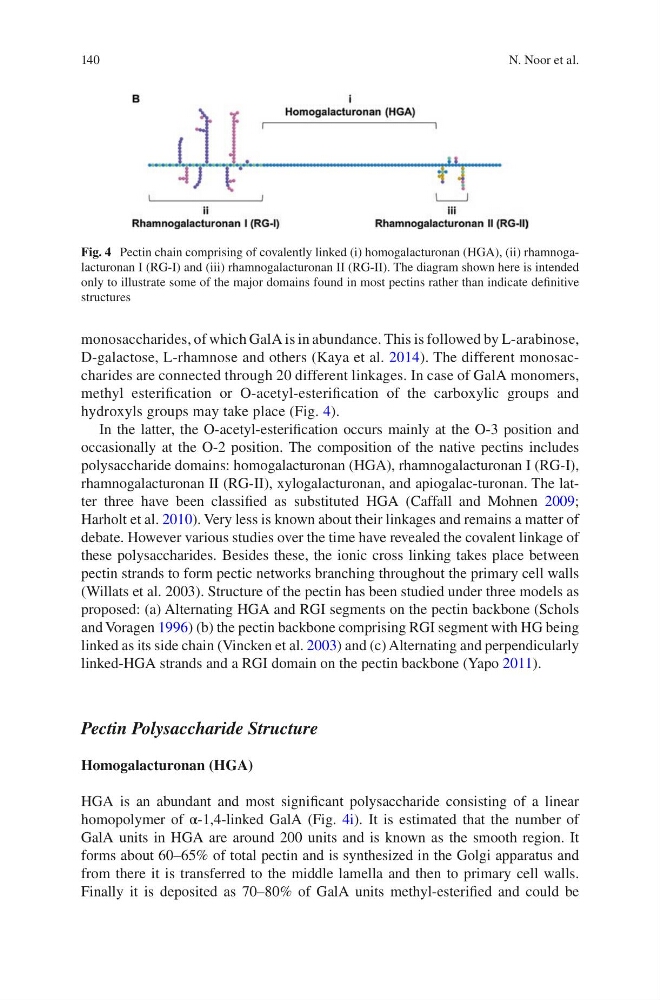
hydroxyls groups may take place (Fig. 4).
In the latter, the O-acetyl-esterification occurs mainly at the O-3 position and
occasionally at the O-2 position. The composition of the native pectins includes
polysaccharide domains: homogalacturonan (HGA), rhamnogalacturonan I (RG-I),
rhamnogalacturonan II (RG-II), xylogalacturonan, and apiogalac-turonan. The latter three have been classified as substituted HGA (Caffall and Mohnen 2009;
Harholt et al. 2010). Very less is known about their linkages and remains a matter of
debate. However various studies over the time have revealed the covalent linkage of
these polysaccharides. Besides these, the ionic cross linking takes place between
pectin strands to form pectic networks branching throughout the primary cell walls
(Willats et al. 2003). Structure of the pectin has been studied under three models as
proposed: (a) Alternating HGA and RGI segments on the pectin backbone (Schols
and Voragen 1996) (b) the pectin backbone comprising RGI segment with HG being
linked as its side chain (Vincken et al. 2003) and (c) Alternating and perpendicularly
linked-HGA strands and a RGI domain on the pectin backbone (Yapo 2011).
Pectin Polysaccharide Structure
Homogalacturonan (HGA)
HGA is an abundant and most significant polysaccharide consisting of a linear
homopolymer of α-1,4-linked GalA (Fig. 4i). It is estimated that the number of
GalA units in HGA are around 200 units and is known as the smooth region. It
forms about 60–65% of total pectin and is synthesized in the Golgi apparatus and
from there it is transferred to the middle lamella and then to primary cell walls.
Finally it is deposited as 70–80% of GalA units methyl-esterified and could be
Fig. 4 Pectin chain comprising of covalently linked (i) homogalacturonan (HGA), (ii) rhamnogalacturonan I (RG-I) and (iii) rhamnogalacturonan II (RG-II). The diagram shown here is intended
only to illustrate some of the major domains found in most pectins rather than indicate definitive
structures
N. Noor et al.
monosaccharides, of which GalA is in abundance. This is followed by L-arabinose,
D-galactose, L-rhamnose and others (Kaya et al. 2014). The different monosaccharides are connected through 20 different linkages. In case of GalA monomers,
methyl esterification or O-acetyl-esterification of the carboxylic groups and
hydroxyls groups may take place (Fig. 4).
In the latter, the O-acetyl-esterification occurs mainly at the O-3 position and
occasionally at the O-2 position. The composition of the native pectins includes
polysaccharide domains: homogalacturonan (HGA), rhamnogalacturonan I (RG-I),
rhamnogalacturonan II (RG-II), xylogalacturonan, and apiogalac-turonan. The latter three have been classified as substituted HGA (Caffall and Mohnen 2009;
Harholt et al. 2010). Very less is known about their linkages and remains a matter of
debate. However various studies over the time have revealed the covalent linkage of
these polysaccharides. Besides these, the ionic cross linking takes place between
pectin strands to form pectic networks branching throughout the primary cell walls
(Willats et al. 2003). Structure of the pectin has been studied under three models as
proposed: (a) Alternating HGA and RGI segments on the pectin backbone (Schols
and Voragen 1996) (b) the pectin backbone comprising RGI segment with HG being
linked as its side chain (Vincken et al. 2003) and (c) Alternating and perpendicularly
linked-HGA strands and a RGI domain on the pectin backbone (Yapo 2011).
Pectin Polysaccharide Structure
Homogalacturonan (HGA)
HGA is an abundant and most significant polysaccharide consisting of a linear
homopolymer of α-1,4-linked GalA (Fig. 4i). It is estimated that the number of
GalA units in HGA are around 200 units and is known as the smooth region. It
forms about 60–65% of total pectin and is synthesized in the Golgi apparatus and
from there it is transferred to the middle lamella and then to primary cell walls.
Finally it is deposited as 70–80% of GalA units methyl-esterified and could be
Fig. 4 Pectin chain comprising of covalently linked (i) homogalacturonan (HGA), (ii) rhamnogalacturonan I (RG-I) and (iii) rhamnogalacturonan II (RG-II). The diagram shown here is intended
only to illustrate some of the major domains found in most pectins rather than indicate definitive
structures
N. Noor et al.