

2.2 Cell Preparation
1. Articular cartilage from femur condyle of young adult bovine.
2. Hank’s Balanced Salt Solution (HBSS) buffer: supplemented
with 100 U/μL penicillin, 100 μg/mL streptomycin, and
2.5 μg/mL fungizone.
3. Culture medium: Dulbecco’s modified culture medium
(DMEM) and Ham’s F12 medium (1:1) supplemented with
10% fetal bovine serum, 50 μg/mL L-ascorbate, 100 U/mL
penicillin, 100 μg/mL streptomycin, and 2.5 μg/mL
fungizone
4. Digestion buffer: 0.25 mg/mL Collagenase type I and
0.25 mg/mL Pronase E in culture medium.
2.3 Bioprinting
Equipment
1. Pneumatic air dispenser.
2. Mechanical pump.
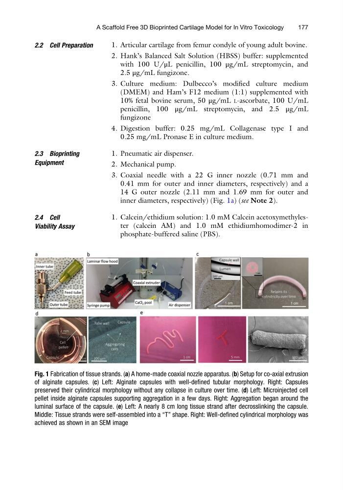
3. Coaxial needle with a 22 G inner nozzle (0.71 mm and
0.41 mm for outer and inner diameters, respectively) and a
14 G outer nozzle (2.11 mm and 1.69 mm for outer and
inner diameters, respectively) (Fig. 1a) (see Note 2).
2.4 Cell
Viability Assay
1. Calcein/ethidium solution: 1.0 mM Calcein acetoxymethylester (calcein AM) and 1.0 mM ethidiumhomodimer-2 in
phosphate-buffered saline (PBS).
Fig. 1 Fabrication of tissue strands. (a) A home-made coaxial nozzle apparatus. (b) Setup for co-axial extrusion
of alginate capsules. (c) Left: Alginate capsules with well-defined tubular morphology. Right: Capsules
preserved their cylindrical morphology without any collapse in culture over time. (d) Left: Microinjected cell
pellet inside alginate capsules supporting aggregation in a few days. Right: Aggregation began around the
luminal surface of the capsule. (e) Left: A nearly 8 cm long tissue strand after decrosslinking the capsule.
Middle: Tissue strands were self-assembled into a “T” shape. Right: Well-defined cylindrical morphology was
achieved as shown in an SEM image
A Scaffold Free 3D Bioprinted Cartilage Model for In Vitro Toxicology
177
1. Articular cartilage from femur condyle of young adult bovine.
2. Hank’s Balanced Salt Solution (HBSS) buffer: supplemented
with 100 U/μL penicillin, 100 μg/mL streptomycin, and
2.5 μg/mL fungizone.
3. Culture medium: Dulbecco’s modified culture medium
(DMEM) and Ham’s F12 medium (1:1) supplemented with
10% fetal bovine serum, 50 μg/mL L-ascorbate, 100 U/mL
penicillin, 100 μg/mL streptomycin, and 2.5 μg/mL
fungizone
4. Digestion buffer: 0.25 mg/mL Collagenase type I and
0.25 mg/mL Pronase E in culture medium.
2.3 Bioprinting
Equipment
1. Pneumatic air dispenser.
2. Mechanical pump.
3. Coaxial needle with a 22 G inner nozzle (0.71 mm and
0.41 mm for outer and inner diameters, respectively) and a
14 G outer nozzle (2.11 mm and 1.69 mm for outer and
inner diameters, respectively) (Fig. 1a) (see Note 2).
2.4 Cell
Viability Assay
1. Calcein/ethidium solution: 1.0 mM Calcein acetoxymethylester (calcein AM) and 1.0 mM ethidiumhomodimer-2 in
phosphate-buffered saline (PBS).
Fig. 1 Fabrication of tissue strands. (a) A home-made coaxial nozzle apparatus. (b) Setup for co-axial extrusion
of alginate capsules. (c) Left: Alginate capsules with well-defined tubular morphology. Right: Capsules
preserved their cylindrical morphology without any collapse in culture over time. (d) Left: Microinjected cell
pellet inside alginate capsules supporting aggregation in a few days. Right: Aggregation began around the
luminal surface of the capsule. (e) Left: A nearly 8 cm long tissue strand after decrosslinking the capsule.
Middle: Tissue strands were self-assembled into a “T” shape. Right: Well-defined cylindrical morphology was
achieved as shown in an SEM image
A Scaffold Free 3D Bioprinted Cartilage Model for In Vitro Toxicology
177