

5. Remove the blocking and permeabilization buffer and apply
the primary antibody solution to the tissue construct.
6. Incubate the tissue construct in the primary antibody solution
overnight at 4
C.
7. After incubation with the primary antibodies, rinse the tissue
construct three times with DPBS for 5 min each.
8. Incubate the tissue construct in the diluted fluorochromeconjugated secondary antibodies for 1 h at room temperature
in the dark.
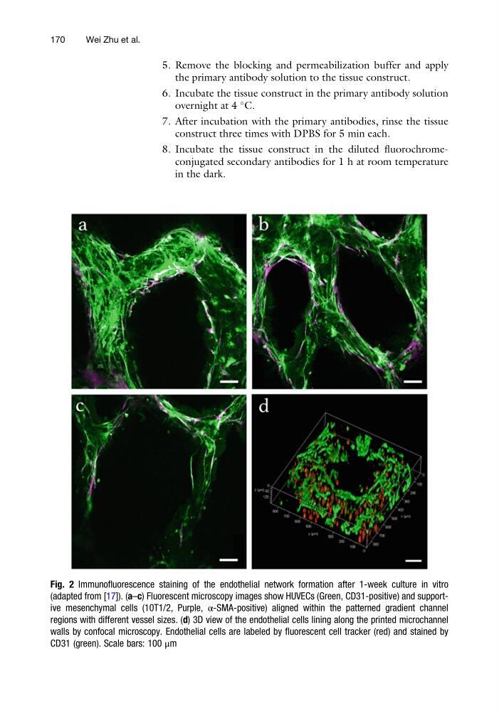
Fig. 2 Immunofluorescence staining of the endothelial network formation after 1-week culture in vitro
(adapted from [17]). (a–c) Fluorescent microscopy images show HUVECs (Green, CD31-positive) and supportive mesenchymal cells (10T1/2, Purple, α-SMA-positive) aligned within the patterned gradient channel
regions with different vessel sizes. (d) 3D view of the endothelial cells lining along the printed microchannel
walls by confocal microscopy. Endothelial cells are labeled by fluorescent cell tracker (red) and stained by
CD31 (green). Scale bars: 100 μm
170
Wei Zhu et al.
the primary antibody solution to the tissue construct.
6. Incubate the tissue construct in the primary antibody solution
overnight at 4
C.
7. After incubation with the primary antibodies, rinse the tissue
construct three times with DPBS for 5 min each.
8. Incubate the tissue construct in the diluted fluorochromeconjugated secondary antibodies for 1 h at room temperature
in the dark.
Fig. 2 Immunofluorescence staining of the endothelial network formation after 1-week culture in vitro
(adapted from [17]). (a–c) Fluorescent microscopy images show HUVECs (Green, CD31-positive) and supportive mesenchymal cells (10T1/2, Purple, α-SMA-positive) aligned within the patterned gradient channel
regions with different vessel sizes. (d) 3D view of the endothelial cells lining along the printed microchannel
walls by confocal microscopy. Endothelial cells are labeled by fluorescent cell tracker (red) and stained by
CD31 (green). Scale bars: 100 μm
170
Wei Zhu et al.