

Structural, Morphological, and Catalytic Properties …
65
d
c
b
a
ν, сm
-1
T, %
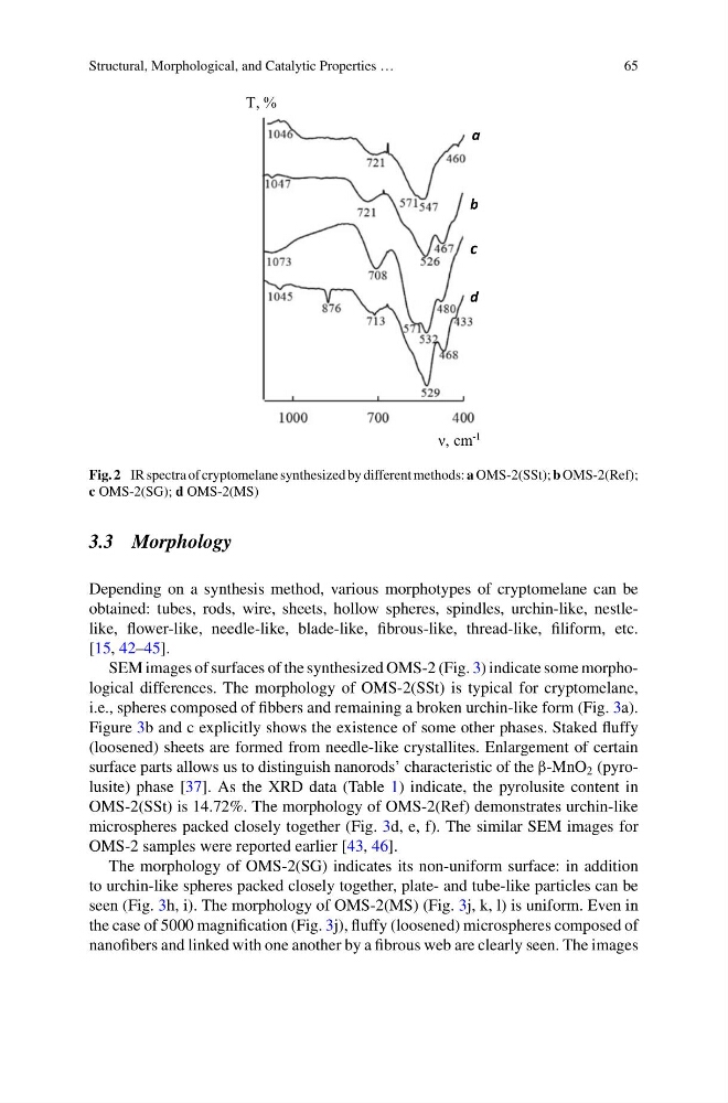
Fig. 2 IR spectra of cryptomelane synthesized by different methods: a OMS-2(SSt); b OMS-2(Ref);
c OMS-2(SG); d OMS-2(MS)
3.3 Morphology
Depending on a synthesis method, various morphotypes of cryptomelane can be
obtained: tubes, rods, wire, sheets, hollow spheres, spindles, urchin-like, nestlelike, flower-like, needle-like, blade-like, fibrous-like, thread-like, filiform, etc.
[15, 42–45].
SEM images of surfaces of the synthesized OMS-2 (Fig. 3) indicate some morphological differences. The morphology of OMS-2(SSt) is typical for cryptomelane,
i.e., spheres composed of fibbers and remaining a broken urchin-like form (Fig. 3a).
Figure 3b and c explicitly shows the existence of some other phases. Staked fluffy
(loosened) sheets are formed from needle-like crystallites. Enlargement of certain
surface parts allows us to distinguish nanorods’ characteristic of the β-MnO 2 (pyrolusite) phase [37]. As the XRD data (Table 1) indicate, the pyrolusite content in
OMS-2(SSt) is 14.72%. The morphology of OMS-2(Ref) demonstrates urchin-like
microspheres packed closely together (Fig. 3d, e, f). The similar SEM images for
OMS-2 samples were reported earlier [43, 46].
The morphology of OMS-2(SG) indicates its non-uniform surface: in addition
to urchin-like spheres packed closely together, plate- and tube-like particles can be
seen (Fig. 3h, i). The morphology of OMS-2(MS) (Fig. 3j, k, l) is uniform. Even in
the case of 5000 magnification (Fig. 3j), fluffy (loosened) microspheres composed of
nanofibers and linked with one another by a fibrous web are clearly seen. The images
65
d
c
b
a
ν, сm
-1
T, %
Fig. 2 IR spectra of cryptomelane synthesized by different methods: a OMS-2(SSt); b OMS-2(Ref);
c OMS-2(SG); d OMS-2(MS)
3.3 Morphology
Depending on a synthesis method, various morphotypes of cryptomelane can be
obtained: tubes, rods, wire, sheets, hollow spheres, spindles, urchin-like, nestlelike, flower-like, needle-like, blade-like, fibrous-like, thread-like, filiform, etc.
[15, 42–45].
SEM images of surfaces of the synthesized OMS-2 (Fig. 3) indicate some morphological differences. The morphology of OMS-2(SSt) is typical for cryptomelane,
i.e., spheres composed of fibbers and remaining a broken urchin-like form (Fig. 3a).
Figure 3b and c explicitly shows the existence of some other phases. Staked fluffy
(loosened) sheets are formed from needle-like crystallites. Enlargement of certain
surface parts allows us to distinguish nanorods’ characteristic of the β-MnO 2 (pyrolusite) phase [37]. As the XRD data (Table 1) indicate, the pyrolusite content in
OMS-2(SSt) is 14.72%. The morphology of OMS-2(Ref) demonstrates urchin-like
microspheres packed closely together (Fig. 3d, e, f). The similar SEM images for
OMS-2 samples were reported earlier [43, 46].
The morphology of OMS-2(SG) indicates its non-uniform surface: in addition
to urchin-like spheres packed closely together, plate- and tube-like particles can be
seen (Fig. 3h, i). The morphology of OMS-2(MS) (Fig. 3j, k, l) is uniform. Even in
the case of 5000 magnification (Fig. 3j), fluffy (loosened) microspheres composed of
nanofibers and linked with one another by a fibrous web are clearly seen. The images