

Physical–Chemical Properties of Magnetite Nanoparticles Doped …
543
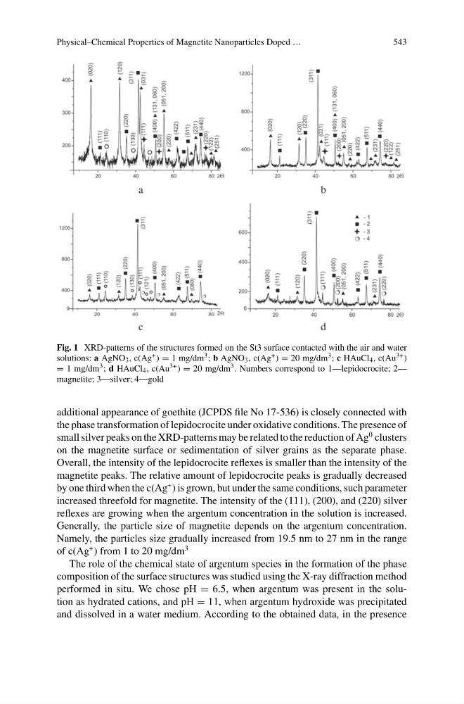
Fig. 1 XRD-patterns of the structures formed on the St3 surface contacted with the air and water
solutions: a AgNO 3 , c(Ag + ) = 1 mg/dm 3 ; b AgNO 3 , c(Ag + ) = 20 mg/dm 3 ; c HAuCl 4 , c(Au 3+ )
= 1 mg/dm 3 ; d HAuCl 4 , c(Au 3+ ) = 20 mg/dm 3 . Numbers correspond to 1—lepidocrocite; 2—
magnetite; 3—silver; 4—gold
additional appearance of goethite (JCPDS file No 17-536) is closely connected with
the phase transformation of lepidocrocite under oxidative conditions. The presence of
small silver peaks on the XRD-patterns may be related to the reduction of Ag
0 clusters
on the magnetite surface or sedimentation of silver grains as the separate phase.
Overall, the intensity of the lepidocrocite reflexes is smaller than the intensity of the
magnetite peaks. The relative amount of lepidocrocite peaks is gradually decreased
by one third when the c(Ag
+ ) is grown, but under the same conditions, such parameter
increased threefold for magnetite. The intensity of the (111), (200), and (220) silver
reflexes are growing when the argentum concentration in the solution is increased.
Generally, the particle size of magnetite depends on the argentum concentration.
Namely, the particles size gradually increased from 19.5 nm to 27 nm in the range
of c(Ag
+ ) from 1 to 20 mg/dm
3
The role of the chemical state of argentum species in the formation of the phase
composition of the surface structures was studied using the X-ray diffraction method
performed in situ. We chose pH = 6.5, when argentum was present in the solution as hydrated cations, and pH = 11, when argentum hydroxide was precipitated
and dissolved in a water medium. According to the obtained data, in the presence
543
Fig. 1 XRD-patterns of the structures formed on the St3 surface contacted with the air and water
solutions: a AgNO 3 , c(Ag + ) = 1 mg/dm 3 ; b AgNO 3 , c(Ag + ) = 20 mg/dm 3 ; c HAuCl 4 , c(Au 3+ )
= 1 mg/dm 3 ; d HAuCl 4 , c(Au 3+ ) = 20 mg/dm 3 . Numbers correspond to 1—lepidocrocite; 2—
magnetite; 3—silver; 4—gold
additional appearance of goethite (JCPDS file No 17-536) is closely connected with
the phase transformation of lepidocrocite under oxidative conditions. The presence of
small silver peaks on the XRD-patterns may be related to the reduction of Ag
0 clusters
on the magnetite surface or sedimentation of silver grains as the separate phase.
Overall, the intensity of the lepidocrocite reflexes is smaller than the intensity of the
magnetite peaks. The relative amount of lepidocrocite peaks is gradually decreased
by one third when the c(Ag
+ ) is grown, but under the same conditions, such parameter
increased threefold for magnetite. The intensity of the (111), (200), and (220) silver
reflexes are growing when the argentum concentration in the solution is increased.
Generally, the particle size of magnetite depends on the argentum concentration.
Namely, the particles size gradually increased from 19.5 nm to 27 nm in the range
of c(Ag
+ ) from 1 to 20 mg/dm
3
The role of the chemical state of argentum species in the formation of the phase
composition of the surface structures was studied using the X-ray diffraction method
performed in situ. We chose pH = 6.5, when argentum was present in the solution as hydrated cations, and pH = 11, when argentum hydroxide was precipitated
and dissolved in a water medium. According to the obtained data, in the presence