

Femtosecond Laser Surface Micro- and Nanotexturing …
245
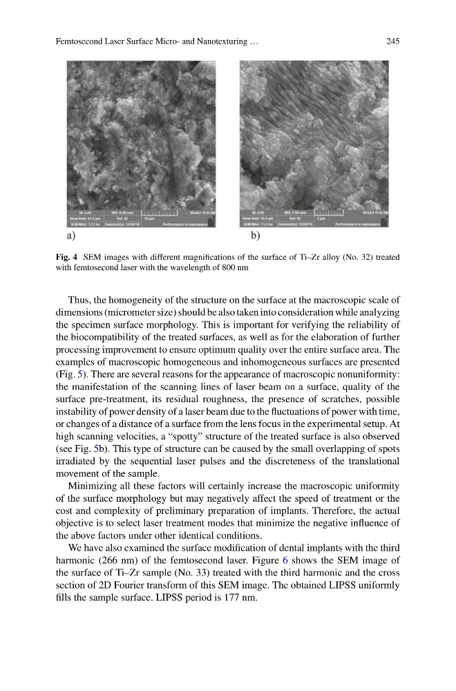
Fig. 4 SEM images with different magnifications of the surface of Ti–Zr alloy (No. 32) treated
with femtosecond laser with the wavelength of 800 nm
Thus, the homogeneity of the structure on the surface at the macroscopic scale of
dimensions (micrometer size) should be also taken into consideration while analyzing
the specimen surface morphology. This is important for verifying the reliability of
the biocompatibility of the treated surfaces, as well as for the elaboration of further
processing improvement to ensure optimum quality over the entire surface area. The
examples of macroscopic homogeneous and inhomogeneous surfaces are presented
(Fig. 5). There are several reasons for the appearance of macroscopic nonuniformity:
the manifestation of the scanning lines of laser beam on a surface, quality of the
surface pre-treatment, its residual roughness, the presence of scratches, possible
instability of power density of a laser beam due to the fluctuations of power with time,
or changes of a distance of a surface from the lens focus in the experimental setup. At
high scanning velocities, a “spotty” structure of the treated surface is also observed
(see Fig. 5b). This type of structure can be caused by the small overlapping of spots
irradiated by the sequential laser pulses and the discreteness of the translational
movement of the sample.
Minimizing all these factors will certainly increase the macroscopic uniformity
of the surface morphology but may negatively affect the speed of treatment or the
cost and complexity of preliminary preparation of implants. Therefore, the actual
objective is to select laser treatment modes that minimize the negative influence of
the above factors under other identical conditions.
We have also examined the surface modification of dental implants with the third
harmonic (266 nm) of the femtosecond laser. Figure 6 shows the SEM image of
the surface of Ti–Zr sample (No. 33) treated with the third harmonic and the cross
section of 2D Fourier transform of this SEM image. The obtained LIPSS uniformly
fills the sample surface. LIPSS period is 177 nm.
245
Fig. 4 SEM images with different magnifications of the surface of Ti–Zr alloy (No. 32) treated
with femtosecond laser with the wavelength of 800 nm
Thus, the homogeneity of the structure on the surface at the macroscopic scale of
dimensions (micrometer size) should be also taken into consideration while analyzing
the specimen surface morphology. This is important for verifying the reliability of
the biocompatibility of the treated surfaces, as well as for the elaboration of further
processing improvement to ensure optimum quality over the entire surface area. The
examples of macroscopic homogeneous and inhomogeneous surfaces are presented
(Fig. 5). There are several reasons for the appearance of macroscopic nonuniformity:
the manifestation of the scanning lines of laser beam on a surface, quality of the
surface pre-treatment, its residual roughness, the presence of scratches, possible
instability of power density of a laser beam due to the fluctuations of power with time,
or changes of a distance of a surface from the lens focus in the experimental setup. At
high scanning velocities, a “spotty” structure of the treated surface is also observed
(see Fig. 5b). This type of structure can be caused by the small overlapping of spots
irradiated by the sequential laser pulses and the discreteness of the translational
movement of the sample.
Minimizing all these factors will certainly increase the macroscopic uniformity
of the surface morphology but may negatively affect the speed of treatment or the
cost and complexity of preliminary preparation of implants. Therefore, the actual
objective is to select laser treatment modes that minimize the negative influence of
the above factors under other identical conditions.
We have also examined the surface modification of dental implants with the third
harmonic (266 nm) of the femtosecond laser. Figure 6 shows the SEM image of
the surface of Ti–Zr sample (No. 33) treated with the third harmonic and the cross
section of 2D Fourier transform of this SEM image. The obtained LIPSS uniformly
fills the sample surface. LIPSS period is 177 nm.