

Femtosecond Laser Surface Micro- and Nanotexturing …
243
in a humidified environment with 5% CO 2 . Cultural media was changed every 3 days
during a 7-day culture period. All experiments were triplicate.
3 Morphology Study of Laser-Treated Surfaces
of Specimens of Dental Type (Zr, Ti–Zr Alloys)
The scanning electron microscopy has been used for the characterization of the topography of the samples of Zr, Ti–Zr alloys, and zirconia ceramics. After femtosecond
laser treatment, almost all specimen surfaces demonstrate the surface periodic
structures.
The commonly accepted mechanism of the laser-induced formation of periodic
surface structures on metals considers the interference of the incident wave and
surface electromagnetic waves (SEW) or surface plasmon polaritons excited at the
metal surface by laser radiation. Thus, the quasiperiodic structures with a period close
to the wavelength of the laser and oriented along the perpendicular to the polarization
plane of the incident light begin to form.
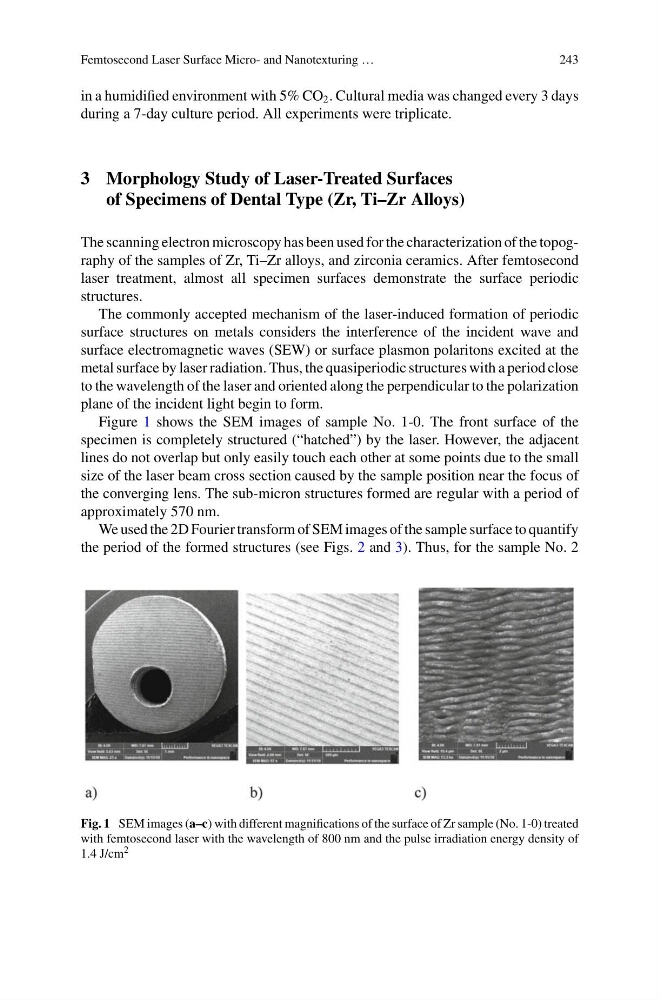
Figure 1 shows the SEM images of sample No. 1-0. The front surface of the
specimen is completely structured (“hatched”) by the laser. However, the adjacent
lines do not overlap but only easily touch each other at some points due to the small
size of the laser beam cross section caused by the sample position near the focus of
the converging lens. The sub-micron structures formed are regular with a period of
approximately 570 nm.
We used the 2D Fourier transform of SEM images of the sample surface to quantify
the period of the formed structures (see Figs. 2 and 3). Thus, for the sample No. 2
Fig. 1 SEM images (a–c) with different magnifications of the surface of Zr sample (No. 1-0) treated
with femtosecond laser with the wavelength of 800 nm and the pulse irradiation energy density of
1.4 J/cm 2
243
in a humidified environment with 5% CO 2 . Cultural media was changed every 3 days
during a 7-day culture period. All experiments were triplicate.
3 Morphology Study of Laser-Treated Surfaces
of Specimens of Dental Type (Zr, Ti–Zr Alloys)
The scanning electron microscopy has been used for the characterization of the topography of the samples of Zr, Ti–Zr alloys, and zirconia ceramics. After femtosecond
laser treatment, almost all specimen surfaces demonstrate the surface periodic
structures.
The commonly accepted mechanism of the laser-induced formation of periodic
surface structures on metals considers the interference of the incident wave and
surface electromagnetic waves (SEW) or surface plasmon polaritons excited at the
metal surface by laser radiation. Thus, the quasiperiodic structures with a period close
to the wavelength of the laser and oriented along the perpendicular to the polarization
plane of the incident light begin to form.
Figure 1 shows the SEM images of sample No. 1-0. The front surface of the
specimen is completely structured (“hatched”) by the laser. However, the adjacent
lines do not overlap but only easily touch each other at some points due to the small
size of the laser beam cross section caused by the sample position near the focus of
the converging lens. The sub-micron structures formed are regular with a period of
approximately 570 nm.
We used the 2D Fourier transform of SEM images of the sample surface to quantify
the period of the formed structures (see Figs. 2 and 3). Thus, for the sample No. 2
Fig. 1 SEM images (a–c) with different magnifications of the surface of Zr sample (No. 1-0) treated
with femtosecond laser with the wavelength of 800 nm and the pulse irradiation energy density of
1.4 J/cm 2