

60
V. Demchenko et al.
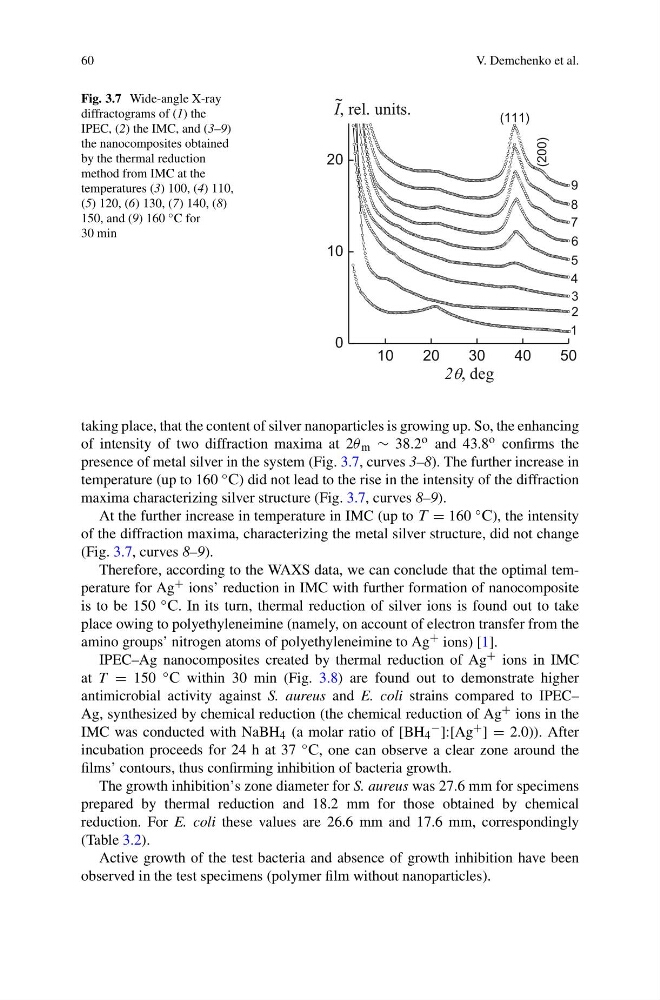
Fig. 3.7 Wide-angle X-ray
diffractograms of (1) the
IPEC, (2) the IMC, and (3–9)
the nanocomposites obtained
by the thermal reduction
method from IMC at the
temperatures (3) 100, (4) 110,
(5) 120, (6) 130, (7) 140, (8)
150, and (9) 160 ◦ C for
30 min
10
20
30
40
50
0
10
20
9
8
7
6
5
4
(200)
(111)
3
2
1
~
I, rel. units.
2q, deg
taking place, that the content of silver nanoparticles is growing up. So, the enhancing
of intensity of two diffraction maxima at 2θ m ∼ 38.2 Ñ and 43.8 Ñ confirms the
presence of metal silver in the system (Fig. 3.7, curves 3–8). The further increase in
temperature (up to 160 ◦ ´) did not lead to the rise in the intensity of the diffraction
maxima characterizing silver structure (Fig. 3.7, curves 8–9).
At the further increase in temperature in IMC (up to µ = 160 ◦ ´), the intensity
of the diffraction maxima, characterizing the metal silver structure, did not change
(Fig. 3.7, curves 8–9).
Therefore, according to the WAXS data, we can conclude that the optimal temperature for Ag + ions’ reduction in IMC with further formation of nanocomposite
is to be 150 ◦ ´. In its turn, thermal reduction of silver ions is found out to take
place owing to polyethyleneimine (namely, on account of electron transfer from the
amino groups’ nitrogen atoms of polyethyleneimine to Ag + ions) [1].
IPEC–Ag nanocomposites created by thermal reduction of Ag + ions in IMC
at T = 150 ◦ ´ within 30 min (Fig. 3.8) are found out to demonstrate higher
antimicrobial activity against S. aureus and E. coli strains compared to IPEC–
Ag, synthesized by chemical reduction (the chemical reduction of Ag + ions in the
IMC was conducted with NaBH 4 (a molar ratio of [BH 4
− ]:[Ag + ] = 2.0)). After
incubation proceeds for 24 h at 37 ◦ ´, one can observe a clear zone around the
films’ contours, thus confirming inhibition of bacteria growth.
The growth inhibition’s zone diameter for S. aureus was 27.6 mm for specimens
prepared by thermal reduction and 18.2 mm for those obtained by chemical
reduction. For E. coli these values are 26.6 mm and 17.6 mm, correspondingly
(Table 3.2).
Active growth of the test bacteria and absence of growth inhibition have been
observed in the test specimens (polymer film without nanoparticles).
V. Demchenko et al.
Fig. 3.7 Wide-angle X-ray
diffractograms of (1) the
IPEC, (2) the IMC, and (3–9)
the nanocomposites obtained
by the thermal reduction
method from IMC at the
temperatures (3) 100, (4) 110,
(5) 120, (6) 130, (7) 140, (8)
150, and (9) 160 ◦ C for
30 min
10
20
30
40
50
0
10
20
9
8
7
6
5
4
(200)
(111)
3
2
1
~
I, rel. units.
2q, deg
taking place, that the content of silver nanoparticles is growing up. So, the enhancing
of intensity of two diffraction maxima at 2θ m ∼ 38.2 Ñ and 43.8 Ñ confirms the
presence of metal silver in the system (Fig. 3.7, curves 3–8). The further increase in
temperature (up to 160 ◦ ´) did not lead to the rise in the intensity of the diffraction
maxima characterizing silver structure (Fig. 3.7, curves 8–9).
At the further increase in temperature in IMC (up to µ = 160 ◦ ´), the intensity
of the diffraction maxima, characterizing the metal silver structure, did not change
(Fig. 3.7, curves 8–9).
Therefore, according to the WAXS data, we can conclude that the optimal temperature for Ag + ions’ reduction in IMC with further formation of nanocomposite
is to be 150 ◦ ´. In its turn, thermal reduction of silver ions is found out to take
place owing to polyethyleneimine (namely, on account of electron transfer from the
amino groups’ nitrogen atoms of polyethyleneimine to Ag + ions) [1].
IPEC–Ag nanocomposites created by thermal reduction of Ag + ions in IMC
at T = 150 ◦ ´ within 30 min (Fig. 3.8) are found out to demonstrate higher
antimicrobial activity against S. aureus and E. coli strains compared to IPEC–
Ag, synthesized by chemical reduction (the chemical reduction of Ag + ions in the
IMC was conducted with NaBH 4 (a molar ratio of [BH 4
− ]:[Ag + ] = 2.0)). After
incubation proceeds for 24 h at 37 ◦ ´, one can observe a clear zone around the
films’ contours, thus confirming inhibition of bacteria growth.
The growth inhibition’s zone diameter for S. aureus was 27.6 mm for specimens
prepared by thermal reduction and 18.2 mm for those obtained by chemical
reduction. For E. coli these values are 26.6 mm and 17.6 mm, correspondingly
(Table 3.2).
Active growth of the test bacteria and absence of growth inhibition have been
observed in the test specimens (polymer film without nanoparticles).