

52
V. Demchenko et al.
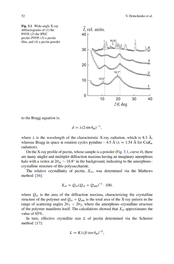
Fig. 3.1 Wide-angle X-ray
diffractograms of (1) the
P4VP, (2) the IPEC
pectin–P4VP, (3) a pectin
film, and (4) a pectin powder
10
20
30
40
0
10
20
30
40
16,8°
19.7°
4
3
2
1
~
I, rel. units.
2q, deg
10.6°
to the Bragg equation is:
d = λ(2 sin θ m )
−1 ,
where λ is the wavelength of the characteristic X-ray radiation, which is 8.3 Å,
whereas Bragg in space at rotation cycles pyridine – 4.5 Å (λ = 1.54 Å for ´uK α
radiation).
On the X-ray profile of pectin, whose sample is a powder (Fig. 3.1, curve 4), there
are many singlet and multiplet diffraction maxima having an imaginary amorphous
halo with a vertex at 2θ m ∼ 16.8 ◦ in the background, indicating to the amorphous–
crystalline structure of this polysaccharide.
The relative crystallinity of pectin, ¸ cr , was determined via the Mathews
method [16]:
X cr = Q cr (Q cr + Q am ) –1 · 100,
where Q cr is the area of the diffraction maxima, characterizing the crystalline
structure of the polymer and Q cr + Q Ãm is the total area of the X-ray pattern in the
range of scattering angles 2θ 1 ÷ 2θ 2 , where the amorphous–crystalline structure
of the polymer manifests itself. The calculations showed that ¸ cr approximates the
value of 65%.
In turn, effective crystallite size L of pectin determined via the Scherrer
method [17]:
L = Kλ(β cos θ m ) –1 ,
V. Demchenko et al.
Fig. 3.1 Wide-angle X-ray
diffractograms of (1) the
P4VP, (2) the IPEC
pectin–P4VP, (3) a pectin
film, and (4) a pectin powder
10
20
30
40
0
10
20
30
40
16,8°
19.7°
4
3
2
1
~
I, rel. units.
2q, deg
10.6°
to the Bragg equation is:
d = λ(2 sin θ m )
−1 ,
where λ is the wavelength of the characteristic X-ray radiation, which is 8.3 Å,
whereas Bragg in space at rotation cycles pyridine – 4.5 Å (λ = 1.54 Å for ´uK α
radiation).
On the X-ray profile of pectin, whose sample is a powder (Fig. 3.1, curve 4), there
are many singlet and multiplet diffraction maxima having an imaginary amorphous
halo with a vertex at 2θ m ∼ 16.8 ◦ in the background, indicating to the amorphous–
crystalline structure of this polysaccharide.
The relative crystallinity of pectin, ¸ cr , was determined via the Mathews
method [16]:
X cr = Q cr (Q cr + Q am ) –1 · 100,
where Q cr is the area of the diffraction maxima, characterizing the crystalline
structure of the polymer and Q cr + Q Ãm is the total area of the X-ray pattern in the
range of scattering angles 2θ 1 ÷ 2θ 2 , where the amorphous–crystalline structure
of the polymer manifests itself. The calculations showed that ¸ cr approximates the
value of 65%.
In turn, effective crystallite size L of pectin determined via the Scherrer
method [17]:
L = Kλ(β cos θ m ) –1 ,