

155
Biomechanical Characterization of Human Red Blood Cells
of the glass surfaces of the chamber were treated with 100 mg/mL bovine
serum albumin (BSA, Sigma, St. Louis, MO, USA) to prevent the beads from
sticking for microbead manipulation experiments (Tan et al. 2009, 2010b).
8.3.2 Experimental Materials Preparation
Fresh blood was drawn from healthy donors by fingertip prick. A small portion of blood was suspended in phosphate-buffered saline (PBS, Sigma) and
then washed three times by centrifugation. A dense RBC sample was obtained
after discarding the top layer of blood after rinsing. In parallel, streptavidin-coated polystyrene beads with a radius of 1.5 μm (Bangs Laboratories,
Fishers, IN) were centrifuged three times in 0.1 mg/mL PBS-BSA solution.
The washed beads were incubated with 1 mg/mL biotin-conjugated concanavalin A (Con A, Sigma) at 4°C for 40 minutes. The beads were then rinsed
three more times in 0.1 mg/mL PBS-BSA and stored in 0.1 mg/mL PBS-BSA
solution with Ca 2+ and Mn 2+ . The prepared polystyrene beads were added to
the RBC suspension and incubated at 25°C for one hour to allow the adhesion
between beads and RBCs. Once the adhesion was confirmed under an optical
microscope, the mixture was diluted in 0.7% hypotonic sodium chloride buffer to allow RBCs to become swollen.
8.3.3 Force Calibration
To acquire the optical trapping force at a certain laser power, it is necessary
to perform force calibration experiments because the trapping force cannot
be measured directly. The usual viscous drag force calibration method was
used here (Henon et al. 1999; Mills et al. 2004; Tan et al. 2010a). A polystyrene bead was trapped at the same separation distance h as that used in cell
stretching experiments. As the chamber was driven to move via the motorized stage, the fluid flow exerted a viscous drag force on the trapped bead.
When the flow velocity increased up to a critical value beyond which the
bead just escaped the laser trap, the bead achieved equilibrium; that is, the
trapping force equaled the viscous drag force. According to Stokes’ law, the
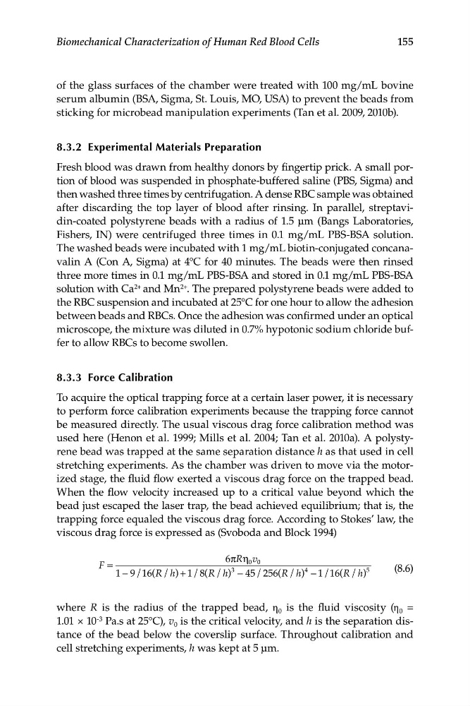
viscous drag force is expressed as (Svoboda and Block 1994)
6π η
R v
0 0
F =
3
4
(8.6)
1 − 9/ 16 (R h
/ ) + 1 /8( R h
/ ) − 45/ 256(R h
/ ) − − 1 1
/ (
6 R h
/ )
5
where R is the radius of the trapped bead, η 0 is the fluid viscosity (η 0 =
1.01 × 10 -3 Pa.s at 25°C), v 0 is the critical velocity, and h is the separation distance of the bead below the coverslip surface. Throughout calibration and
cell stretching experiments, h was kept at 5 μm.
Biomechanical Characterization of Human Red Blood Cells
of the glass surfaces of the chamber were treated with 100 mg/mL bovine
serum albumin (BSA, Sigma, St. Louis, MO, USA) to prevent the beads from
sticking for microbead manipulation experiments (Tan et al. 2009, 2010b).
8.3.2 Experimental Materials Preparation
Fresh blood was drawn from healthy donors by fingertip prick. A small portion of blood was suspended in phosphate-buffered saline (PBS, Sigma) and
then washed three times by centrifugation. A dense RBC sample was obtained
after discarding the top layer of blood after rinsing. In parallel, streptavidin-coated polystyrene beads with a radius of 1.5 μm (Bangs Laboratories,
Fishers, IN) were centrifuged three times in 0.1 mg/mL PBS-BSA solution.
The washed beads were incubated with 1 mg/mL biotin-conjugated concanavalin A (Con A, Sigma) at 4°C for 40 minutes. The beads were then rinsed
three more times in 0.1 mg/mL PBS-BSA and stored in 0.1 mg/mL PBS-BSA
solution with Ca 2+ and Mn 2+ . The prepared polystyrene beads were added to
the RBC suspension and incubated at 25°C for one hour to allow the adhesion
between beads and RBCs. Once the adhesion was confirmed under an optical
microscope, the mixture was diluted in 0.7% hypotonic sodium chloride buffer to allow RBCs to become swollen.
8.3.3 Force Calibration
To acquire the optical trapping force at a certain laser power, it is necessary
to perform force calibration experiments because the trapping force cannot
be measured directly. The usual viscous drag force calibration method was
used here (Henon et al. 1999; Mills et al. 2004; Tan et al. 2010a). A polystyrene bead was trapped at the same separation distance h as that used in cell
stretching experiments. As the chamber was driven to move via the motorized stage, the fluid flow exerted a viscous drag force on the trapped bead.
When the flow velocity increased up to a critical value beyond which the
bead just escaped the laser trap, the bead achieved equilibrium; that is, the
trapping force equaled the viscous drag force. According to Stokes’ law, the
viscous drag force is expressed as (Svoboda and Block 1994)
6π η
R v
0 0
F =
3
4
(8.6)
1 − 9/ 16 (R h
/ ) + 1 /8( R h
/ ) − 45/ 256(R h
/ ) − − 1 1
/ (
6 R h
/ )
5
where R is the radius of the trapped bead, η 0 is the fluid viscosity (η 0 =
1.01 × 10 -3 Pa.s at 25°C), v 0 is the critical velocity, and h is the separation distance of the bead below the coverslip surface. Throughout calibration and
cell stretching experiments, h was kept at 5 μm.