

9.1 Introduction
167
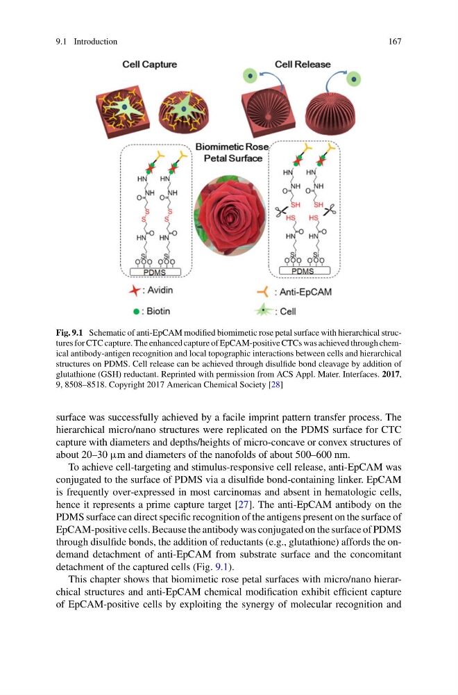
Fig. 9.1 Schematic of anti-EpCAM modified biomimetic rose petal surface with hierarchical structures for CTC capture. The enhanced capture of EpCAM-positive CTCs was achieved through chemical antibody-antigen recognition and local topographic interactions between cells and hierarchical
structures on PDMS. Cell release can be achieved through disulfide bond cleavage by addition of
glutathione (GSH) reductant. Reprinted with permission from ACS Appl. Mater. Interfaces. 2017,
9, 8508–8518. Copyright 2017 American Chemical Society [28]
surface was successfully achieved by a facile imprint pattern transfer process. The
hierarchical micro/nano structures were replicated on the PDMS surface for CTC
capture with diameters and depths/heights of micro-concave or convex structures of
about 20–30 μm and diameters of the nanofolds of about 500–600 nm.
To achieve cell-targeting and stimulus-responsive cell release, anti-EpCAM was
conjugated to the surface of PDMS via a disulfide bond-containing linker. EpCAM
is frequently over-expressed in most carcinomas and absent in hematologic cells,
hence it represents a prime capture target [27]. The anti-EpCAM antibody on the
PDMS surface can direct specific recognition of the antigens present on the surface of
EpCAM-positive cells. Because the antibody was conjugated on the surface of PDMS
through disulfide bonds, the addition of reductants (e.g., glutathione) affords the ondemand detachment of anti-EpCAM from substrate surface and the concomitant
detachment of the captured cells (Fig. 9.1).
This chapter shows that biomimetic rose petal surfaces with micro/nano hierarchical structures and anti-EpCAM chemical modification exhibit efficient capture
of EpCAM-positive cells by exploiting the synergy of molecular recognition and
167
Fig. 9.1 Schematic of anti-EpCAM modified biomimetic rose petal surface with hierarchical structures for CTC capture. The enhanced capture of EpCAM-positive CTCs was achieved through chemical antibody-antigen recognition and local topographic interactions between cells and hierarchical
structures on PDMS. Cell release can be achieved through disulfide bond cleavage by addition of
glutathione (GSH) reductant. Reprinted with permission from ACS Appl. Mater. Interfaces. 2017,
9, 8508–8518. Copyright 2017 American Chemical Society [28]
surface was successfully achieved by a facile imprint pattern transfer process. The
hierarchical micro/nano structures were replicated on the PDMS surface for CTC
capture with diameters and depths/heights of micro-concave or convex structures of
about 20–30 μm and diameters of the nanofolds of about 500–600 nm.
To achieve cell-targeting and stimulus-responsive cell release, anti-EpCAM was
conjugated to the surface of PDMS via a disulfide bond-containing linker. EpCAM
is frequently over-expressed in most carcinomas and absent in hematologic cells,
hence it represents a prime capture target [27]. The anti-EpCAM antibody on the
PDMS surface can direct specific recognition of the antigens present on the surface of
EpCAM-positive cells. Because the antibody was conjugated on the surface of PDMS
through disulfide bonds, the addition of reductants (e.g., glutathione) affords the ondemand detachment of anti-EpCAM from substrate surface and the concomitant
detachment of the captured cells (Fig. 9.1).
This chapter shows that biomimetic rose petal surfaces with micro/nano hierarchical structures and anti-EpCAM chemical modification exhibit efficient capture
of EpCAM-positive cells by exploiting the synergy of molecular recognition and