

2.4 Methods and Materials
51
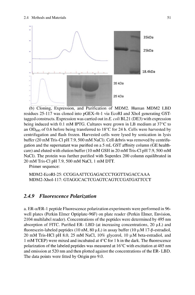
(b) Cloning, Expression, and Purification of MDM2. Human MDM2 LBD
residues 25-117 was cloned into pGEX-4t-1 via EcoRI and XhoI generating GSTtagged constructs. Expression was carried out in E. coli BL21 (DE3) with expression
being induced with 0.1 mM IPTG. Cultures were grown in LB medium at 37°C to
an OD 600 of 0.6 before being transferred to 18°C for 24 h. Cells were harvested by
centrifugation and flash frozen. Harvested cells were lysed by sonication in lysis
buffer (20 mM Tris-Cl pH 7.9, 500 mM NaCl). Cell debris was removed by centrifugation and the supernatant was purified on a 5 mL GST affinity column (GE healthcare) and eluted with elution buffer (10 mM GSH in 20 mM Tris-Cl pH 7.9, 500 mM
NaCl). The protein was further purified with Superdex 200 column equilibrated in
20 mM Tris-Cl pH 7.9, 500 mM NaCl, 1 mM DTT.
Primer sequence:
MDM2-EcoRI-25: CCGGAATTCGAGACCCTGGTTAGACCAAA
MDM2-XhoI-117: GTAGGCACTCGAGTCAGTCCGATGATTCCT
2.4.9 Fluorescence Polarization
a. ER-α/ER-1 peptide Fluorescence polarization experiments were performed in 96well plates (Perkin Elmer Optiplate-96F) on plate reader (Perkin Elmer, Envision,
2104 multilabel reader). Concentrations of the peptides were determined by 495 nm
absorption of FITC. Purified ER- LBD (at increasing concentrations, 20 μL) and
fluorescein-labeled peptides (10 nM, 80 μL) in assay buffer (10 μM 17-β-estradiol,
20 mM Tris-HCl pH 8.0, 25 mM NaCl, 10% glycerol, 10 μM beta-estradiol, and
1 mM TCEP) were mixed and incubated at 4°C for 1 h in the dark. The fluorescence
polarization of the labeled peptides was measured at 16°C with excitation at 485 nm
and emission at 520 nm and then plotted against the concentrations of the ER- LBD.
The data points were fitted by Origin pro 9.0.
51
(b) Cloning, Expression, and Purification of MDM2. Human MDM2 LBD
residues 25-117 was cloned into pGEX-4t-1 via EcoRI and XhoI generating GSTtagged constructs. Expression was carried out in E. coli BL21 (DE3) with expression
being induced with 0.1 mM IPTG. Cultures were grown in LB medium at 37°C to
an OD 600 of 0.6 before being transferred to 18°C for 24 h. Cells were harvested by
centrifugation and flash frozen. Harvested cells were lysed by sonication in lysis
buffer (20 mM Tris-Cl pH 7.9, 500 mM NaCl). Cell debris was removed by centrifugation and the supernatant was purified on a 5 mL GST affinity column (GE healthcare) and eluted with elution buffer (10 mM GSH in 20 mM Tris-Cl pH 7.9, 500 mM
NaCl). The protein was further purified with Superdex 200 column equilibrated in
20 mM Tris-Cl pH 7.9, 500 mM NaCl, 1 mM DTT.
Primer sequence:
MDM2-EcoRI-25: CCGGAATTCGAGACCCTGGTTAGACCAAA
MDM2-XhoI-117: GTAGGCACTCGAGTCAGTCCGATGATTCCT
2.4.9 Fluorescence Polarization
a. ER-α/ER-1 peptide Fluorescence polarization experiments were performed in 96well plates (Perkin Elmer Optiplate-96F) on plate reader (Perkin Elmer, Envision,
2104 multilabel reader). Concentrations of the peptides were determined by 495 nm
absorption of FITC. Purified ER- LBD (at increasing concentrations, 20 μL) and
fluorescein-labeled peptides (10 nM, 80 μL) in assay buffer (10 μM 17-β-estradiol,
20 mM Tris-HCl pH 8.0, 25 mM NaCl, 10% glycerol, 10 μM beta-estradiol, and
1 mM TCEP) were mixed and incubated at 4°C for 1 h in the dark. The fluorescence
polarization of the labeled peptides was measured at 16°C with excitation at 485 nm
and emission at 520 nm and then plotted against the concentrations of the ER- LBD.
The data points were fitted by Origin pro 9.0.