

69
3.4.2 Nanomaterials and Alternative Models of the Placenta
The studies on the transport across the perfused placenta documented that polystyrene nanoparticles accumulated in the syncytiotrophoblast through an energydependent mechanism. The transport across the barrier was polarized, being more
rapid from the maternal to the fetal environment than in the opposite direction. The
accumulation of nanomaterials inside the placenta did not affect the viability of the
explants, and only the nanomaterials with basic functionalization caused fluid leakage (Grafmueller et al. 2015a, 2015b; Wick et al. 2010). However, in vitro exposure
of trophoblast cells to acidic polystyrene nanoparticles revealed a certain grade of
cytotoxicity, as increased expression of apoptosis markers (Huang et al. 2015). The
same is true for exposure to copper nanoparticles of an immortalized trophoblat cell
line, in which the compound induced apoptosis, necrosis, and arrest of the cell
cycle. The IC50 after 48 and 72 hours of exposure was assessed at 20 and 10 μg/ml
(Zhang et al. 2018). Silica and gold nanoparticles accumulated in the trophoblast or
in the perfused placenta. However, only the former crossed the barrier by 5%, and
the latter ones did not reach the fetal environment during the observational time of
6 hours. Studies in cells cultured in vitro confirmed these results (Myllynen et al.
2008; Poulsen et al. 2015). Polystyrene nanoparticles crossed the BeWo cells in
TransWell® A without signs of cytotoxicity (Cartwright et al. 2012). Other nanoparticles tested with the TransWell® methods involved the clinical exploitation of new
materials.
The super paramagnetic nanoparticles of iron oxide are powerful probes approved
for magnetic resonance imaging; the cobalt and chromium alloy nanoparticles can
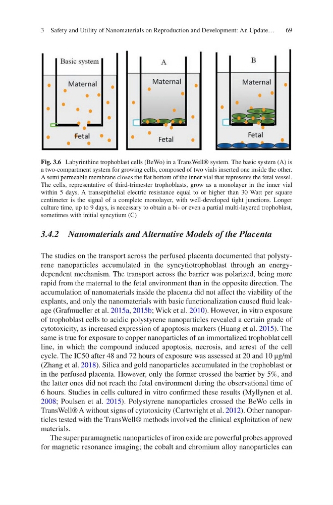
Fig. 3.6 Labyrinthine trophoblast cells (BeWo) in a TransWell® system. The basic system (A) is
a two-compartment system for growing cells, composed of two vials inserted one inside the other.
A semi permeable membrane closes the flat bottom of the inner vial that represents the fetal vessel.
The cells, representative of third-trimester trophoblasts, grow as a monolayer in the inner vial
within 5 days. A transepithelial electric resistance equal to or higher than 30 Watt per square
centimeter is the signal of a complete monolayer, with well-developed tight junctions. Longer
culture time, up to 9 days, is necessary to obtain a bi- or even a partial multi-layered trophoblast,
sometimes with initial syncytium (C)
3 Safety and Utility of Nanomaterials on Reproduction and Development: An Update…
3.4.2 Nanomaterials and Alternative Models of the Placenta
The studies on the transport across the perfused placenta documented that polystyrene nanoparticles accumulated in the syncytiotrophoblast through an energydependent mechanism. The transport across the barrier was polarized, being more
rapid from the maternal to the fetal environment than in the opposite direction. The
accumulation of nanomaterials inside the placenta did not affect the viability of the
explants, and only the nanomaterials with basic functionalization caused fluid leakage (Grafmueller et al. 2015a, 2015b; Wick et al. 2010). However, in vitro exposure
of trophoblast cells to acidic polystyrene nanoparticles revealed a certain grade of
cytotoxicity, as increased expression of apoptosis markers (Huang et al. 2015). The
same is true for exposure to copper nanoparticles of an immortalized trophoblat cell
line, in which the compound induced apoptosis, necrosis, and arrest of the cell
cycle. The IC50 after 48 and 72 hours of exposure was assessed at 20 and 10 μg/ml
(Zhang et al. 2018). Silica and gold nanoparticles accumulated in the trophoblast or
in the perfused placenta. However, only the former crossed the barrier by 5%, and
the latter ones did not reach the fetal environment during the observational time of
6 hours. Studies in cells cultured in vitro confirmed these results (Myllynen et al.
2008; Poulsen et al. 2015). Polystyrene nanoparticles crossed the BeWo cells in
TransWell® A without signs of cytotoxicity (Cartwright et al. 2012). Other nanoparticles tested with the TransWell® methods involved the clinical exploitation of new
materials.
The super paramagnetic nanoparticles of iron oxide are powerful probes approved
for magnetic resonance imaging; the cobalt and chromium alloy nanoparticles can
Fig. 3.6 Labyrinthine trophoblast cells (BeWo) in a TransWell® system. The basic system (A) is
a two-compartment system for growing cells, composed of two vials inserted one inside the other.
A semi permeable membrane closes the flat bottom of the inner vial that represents the fetal vessel.
The cells, representative of third-trimester trophoblasts, grow as a monolayer in the inner vial
within 5 days. A transepithelial electric resistance equal to or higher than 30 Watt per square
centimeter is the signal of a complete monolayer, with well-developed tight junctions. Longer
culture time, up to 9 days, is necessary to obtain a bi- or even a partial multi-layered trophoblast,
sometimes with initial syncytium (C)
3 Safety and Utility of Nanomaterials on Reproduction and Development: An Update…