

211
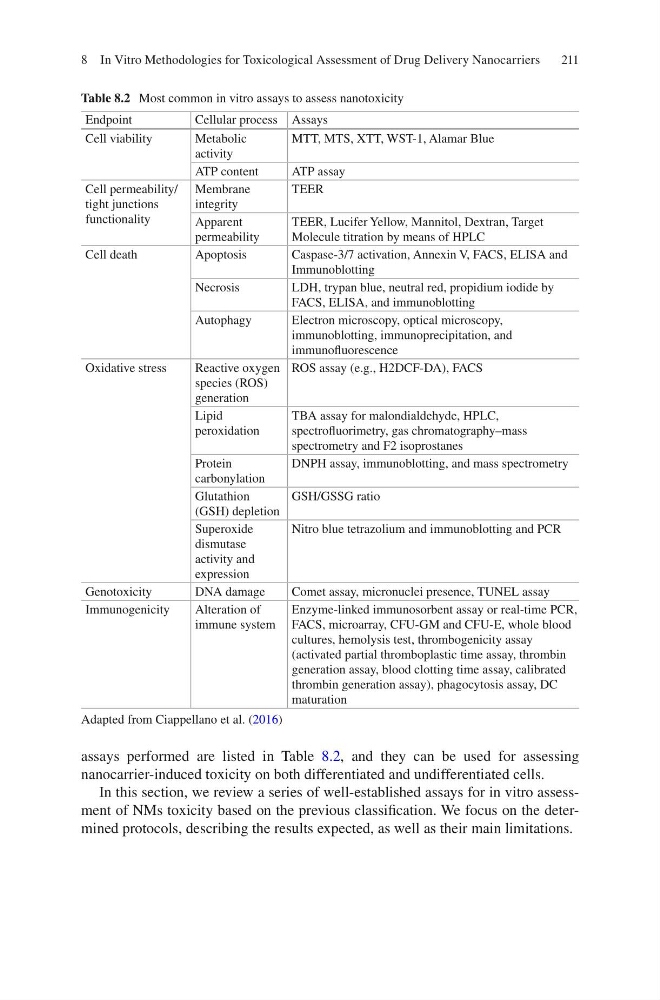
assays performed are listed in Table 8.2, and they can be used for assessing
nanocarrier- induced toxicity on both differentiated and undifferentiated cells.
In this section, we review a series of well-established assays for in vitro assessment of NMs toxicity based on the previous classification. We focus on the determined protocols, describing the results expected, as well as their main limitations.
Table 8.2 Most common in vitro assays to assess nanotoxicity
Endpoint
Cellular process Assays
Cell viability
Metabolic
activity
MTT, MTS, XTT, WST-1, Alamar Blue
ATP content
ATP assay
Cell permeability/
tight junctions
functionality
Membrane
integrity
TEER
Apparent
permeability
TEER, Lucifer Yellow, Mannitol, Dextran, Target
Molecule titration by means of HPLC
Cell death
Apoptosis
Caspase-3/7 activation, Annexin V, FACS, ELISA and
Immunoblotting
Necrosis
LDH, trypan blue, neutral red, propidium iodide by
FACS, ELISA, and immunoblotting
Autophagy
Electron microscopy, optical microscopy,
immunoblotting, immunoprecipitation, and
immunofluorescence
Oxidative stress
Reactive oxygen
species (ROS)
generation
ROS assay (e.g., H2DCF-DA), FACS
Lipid
peroxidation
TBA assay for malondialdehyde, HPLC,
spectrofluorimetry, gas chromatography–mass
spectrometry and F2 isoprostanes
Protein
carbonylation
DNPH assay, immunoblotting, and mass spectrometry
Glutathion
(GSH) depletion
GSH/GSSG ratio
Superoxide
dismutase
activity and
expression
Nitro blue tetrazolium and immunoblotting and PCR
Genotoxicity
DNA damage
Comet assay, micronuclei presence, TUNEL assay
Immunogenicity
Alteration of
immune system
Enzyme-linked immunosorbent assay or real-time PCR,
FACS, microarray, CFU-GM and CFU-E, whole blood
cultures, hemolysis test, thrombogenicity assay
(activated partial thromboplastic time assay, thrombin
generation assay, blood clotting time assay, calibrated
thrombin generation assay), phagocytosis assay, DC
maturation
Adapted from Ciappellano et al. (2016)
8 In Vitro Methodologies for Toxicological Assessment of Drug Delivery Nanocarriers
assays performed are listed in Table 8.2, and they can be used for assessing
nanocarrier- induced toxicity on both differentiated and undifferentiated cells.
In this section, we review a series of well-established assays for in vitro assessment of NMs toxicity based on the previous classification. We focus on the determined protocols, describing the results expected, as well as their main limitations.
Table 8.2 Most common in vitro assays to assess nanotoxicity
Endpoint
Cellular process Assays
Cell viability
Metabolic
activity
MTT, MTS, XTT, WST-1, Alamar Blue
ATP content
ATP assay
Cell permeability/
tight junctions
functionality
Membrane
integrity
TEER
Apparent
permeability
TEER, Lucifer Yellow, Mannitol, Dextran, Target
Molecule titration by means of HPLC
Cell death
Apoptosis
Caspase-3/7 activation, Annexin V, FACS, ELISA and
Immunoblotting
Necrosis
LDH, trypan blue, neutral red, propidium iodide by
FACS, ELISA, and immunoblotting
Autophagy
Electron microscopy, optical microscopy,
immunoblotting, immunoprecipitation, and
immunofluorescence
Oxidative stress
Reactive oxygen
species (ROS)
generation
ROS assay (e.g., H2DCF-DA), FACS
Lipid
peroxidation
TBA assay for malondialdehyde, HPLC,
spectrofluorimetry, gas chromatography–mass
spectrometry and F2 isoprostanes
Protein
carbonylation
DNPH assay, immunoblotting, and mass spectrometry
Glutathion
(GSH) depletion
GSH/GSSG ratio
Superoxide
dismutase
activity and
expression
Nitro blue tetrazolium and immunoblotting and PCR
Genotoxicity
DNA damage
Comet assay, micronuclei presence, TUNEL assay
Immunogenicity
Alteration of
immune system
Enzyme-linked immunosorbent assay or real-time PCR,
FACS, microarray, CFU-GM and CFU-E, whole blood
cultures, hemolysis test, thrombogenicity assay
(activated partial thromboplastic time assay, thrombin
generation assay, blood clotting time assay, calibrated
thrombin generation assay), phagocytosis assay, DC
maturation
Adapted from Ciappellano et al. (2016)
8 In Vitro Methodologies for Toxicological Assessment of Drug Delivery Nanocarriers