

4
Results from these studies showed that soluble nanoparticles had the possibility
to have toxicity to all organisms. Many research studied proved that the adverse
effects usually come from ions released from cobalt, silver, and some other nanoparticles. To examine the toxic properties of nanomaterials, one needs to investigate
where toxicity is found (mainly research laboratories and industry) and which ion
interacts with. This includes the investigation of the absorption of nanoparticles by
living cells, soils and organic material, and the impact of nanoparticle deposition in
the environment. Nanomaterial interactions and impacts must be understood before
scientists and the U.S. Food and Drug Administration approve the use of these
materials.
Nanomaterial toxicity can be classified based on the immense research and studies
of nanotoxicants. They can further be identified as a hazard by means of ecotoxicological data, as well as through their physical–chemical properties,
biodegradability, accumulation, particle size, and composition. The four main
sources of nanoparticle release whereby the ecosystem is contaminated are
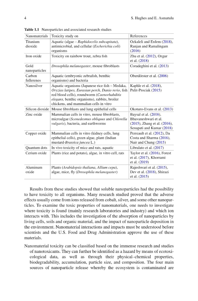
Table 1.1 Nanoparticles and associated research studies
Nanomaterials Toxicity study on
References
Titanium
dioxide
Aquatic (algae – Raphidocelis subcapitata),
antimicrobial, and cellular (Escherichia coli)
organisms
Ozkaleli and Erdem (2018),
Ranjan and Ramalingam
(2016)
Iron oxide
Toxicity on rainbow trout, zebra fish
Zhu et al. (2012), Ozgur
et al. (2018)
Gold
nanoparticles
Drosophila melanogaster, mouse fibroblasts
Coradeghini et al. (2013)
Carbon
fullerenes
Aquatic (embryonic zebrafish, benthic
organisms) and bacteria
Oberdörster et al. (2006)
Nanosilver
Aquatic organisms (Japanese rice fish – Medaka,
Oryzias latipes, Eurasian perch, Danio rerio, fish
red blood cells), roundworm (Caenorhabditis
elegans, benthic organisms), rabbits, broiler
chickens, and mammalian cells in vitro
Kaphle et al. (2018),
Pulit-Prociak (2015)
Silicon dioxide Mouse fibroblasts and lung epithelial cells
Okoturo-Evans et al. (2013)
Zinc oxide
Mammalian cells in vitro, mouse fibroblasts,
microalgae (Scenedesmus obliquus and Chlorella
vulgaris), bacteria, and earthworms
Baysal et al. (2018),
Bhuvaneshwari et al.
(2015), Zhang et al. (2016),
Senapati and Kumar (2018)
Copper oxide Mammalian cells in vitro (kidney cells, lung
epithelial cells), green algae, plant (Indian
mustard-Brassica juncea L.)
Perreault et al. (2012), Da
Costa and Sharma (2016),
Nair and Chung (2015)
Quantum dots In vivo toxicity of mice and rats, aquatic
Libralato et al. (2017)
Cerium oxide Plants (rice and potato), algae, in vitro cell, rats
Taylor et al. (2016), Forest
et al. (2017), Khorrami
et al. (2019)
Aluminum
oxide
Plants (Arabidopsis thaliana, Allium cepa),
algae, mice, fly (Drosophila melanogaster)
Rajeshwari et al. (2015),
Dev et al. (2018), Shirazi
et al. (2015)
S. Hughes and E. Asmatulu
Results from these studies showed that soluble nanoparticles had the possibility
to have toxicity to all organisms. Many research studied proved that the adverse
effects usually come from ions released from cobalt, silver, and some other nanoparticles. To examine the toxic properties of nanomaterials, one needs to investigate
where toxicity is found (mainly research laboratories and industry) and which ion
interacts with. This includes the investigation of the absorption of nanoparticles by
living cells, soils and organic material, and the impact of nanoparticle deposition in
the environment. Nanomaterial interactions and impacts must be understood before
scientists and the U.S. Food and Drug Administration approve the use of these
materials.
Nanomaterial toxicity can be classified based on the immense research and studies
of nanotoxicants. They can further be identified as a hazard by means of ecotoxicological data, as well as through their physical–chemical properties,
biodegradability, accumulation, particle size, and composition. The four main
sources of nanoparticle release whereby the ecosystem is contaminated are
Table 1.1 Nanoparticles and associated research studies
Nanomaterials Toxicity study on
References
Titanium
dioxide
Aquatic (algae – Raphidocelis subcapitata),
antimicrobial, and cellular (Escherichia coli)
organisms
Ozkaleli and Erdem (2018),
Ranjan and Ramalingam
(2016)
Iron oxide
Toxicity on rainbow trout, zebra fish
Zhu et al. (2012), Ozgur
et al. (2018)
Gold
nanoparticles
Drosophila melanogaster, mouse fibroblasts
Coradeghini et al. (2013)
Carbon
fullerenes
Aquatic (embryonic zebrafish, benthic
organisms) and bacteria
Oberdörster et al. (2006)
Nanosilver
Aquatic organisms (Japanese rice fish – Medaka,
Oryzias latipes, Eurasian perch, Danio rerio, fish
red blood cells), roundworm (Caenorhabditis
elegans, benthic organisms), rabbits, broiler
chickens, and mammalian cells in vitro
Kaphle et al. (2018),
Pulit-Prociak (2015)
Silicon dioxide Mouse fibroblasts and lung epithelial cells
Okoturo-Evans et al. (2013)
Zinc oxide
Mammalian cells in vitro, mouse fibroblasts,
microalgae (Scenedesmus obliquus and Chlorella
vulgaris), bacteria, and earthworms
Baysal et al. (2018),
Bhuvaneshwari et al.
(2015), Zhang et al. (2016),
Senapati and Kumar (2018)
Copper oxide Mammalian cells in vitro (kidney cells, lung
epithelial cells), green algae, plant (Indian
mustard-Brassica juncea L.)
Perreault et al. (2012), Da
Costa and Sharma (2016),
Nair and Chung (2015)
Quantum dots In vivo toxicity of mice and rats, aquatic
Libralato et al. (2017)
Cerium oxide Plants (rice and potato), algae, in vitro cell, rats
Taylor et al. (2016), Forest
et al. (2017), Khorrami
et al. (2019)
Aluminum
oxide
Plants (Arabidopsis thaliana, Allium cepa),
algae, mice, fly (Drosophila melanogaster)
Rajeshwari et al. (2015),
Dev et al. (2018), Shirazi
et al. (2015)
S. Hughes and E. Asmatulu