

200
OPTICAL AND VIBRATIONAL SPECTROSCOPY
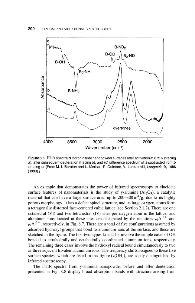
Figure 8.5. FTIR spectra of boron nitride nanopowder surfaces after activation at 875 K (tracing
a), after subsequent deuteration (tracing b), and (c) difference spectrum of a subtracted from b
(tracing c). [From M.4. Baraton and L. Merhari, P. Quintard, V. Lorezenvilli, Langrnuir, 9, 1486
(1 993).]
An example that demonstrates the power of infrared spectroscopy to elucidate
surface features of nanomaterials is the study of y-alumina (A1203), a catalytic
material that can have a large surface area, up to 20&300m2/g, due to its highly
porous morphology. It has a defect spinel structure, and its large oxygen atoms form
a tetragonally distorted face-centered cubic lattice (see Section 2.1.2). There are one
octahedral (VI) and two tetrahedral (IV) sites per oxygen atom in the lattice, and
aluminum ions located at these sites are designated by the notations vIA13+ and
IvA13+, respectively, in Fig. 8.7. There are a total of five configurations assumed by
adsorbed hydroxyl groups that bond to aluminum ions at the surface, and these are
sketched in the figure. The first two, types Ia and Ib, involve the simple cases of OH
bonded to tetrahedrally and octahedrally coordinated aluminum ions, respectively.
The remaining three cases involve the hydroxyl radical bound simultaneously to two
or three adjacent trivalent aluminum ions. The frequency shifts assigned to these five
surface species, which are listed in the figure (v(OH)), are easily distinguished by
infrared spectroscopy.
The FTIR spectra from y-alumina nanopowder before and after deuteration
presented in Fig. 8.8 display broad absorption bands with structure arising from
OPTICAL AND VIBRATIONAL SPECTROSCOPY
Figure 8.5. FTIR spectra of boron nitride nanopowder surfaces after activation at 875 K (tracing
a), after subsequent deuteration (tracing b), and (c) difference spectrum of a subtracted from b
(tracing c). [From M.4. Baraton and L. Merhari, P. Quintard, V. Lorezenvilli, Langrnuir, 9, 1486
(1 993).]
An example that demonstrates the power of infrared spectroscopy to elucidate
surface features of nanomaterials is the study of y-alumina (A1203), a catalytic
material that can have a large surface area, up to 20&300m2/g, due to its highly
porous morphology. It has a defect spinel structure, and its large oxygen atoms form
a tetragonally distorted face-centered cubic lattice (see Section 2.1.2). There are one
octahedral (VI) and two tetrahedral (IV) sites per oxygen atom in the lattice, and
aluminum ions located at these sites are designated by the notations vIA13+ and
IvA13+, respectively, in Fig. 8.7. There are a total of five configurations assumed by
adsorbed hydroxyl groups that bond to aluminum ions at the surface, and these are
sketched in the figure. The first two, types Ia and Ib, involve the simple cases of OH
bonded to tetrahedrally and octahedrally coordinated aluminum ions, respectively.
The remaining three cases involve the hydroxyl radical bound simultaneously to two
or three adjacent trivalent aluminum ions. The frequency shifts assigned to these five
surface species, which are listed in the figure (v(OH)), are easily distinguished by
infrared spectroscopy.
The FTIR spectra from y-alumina nanopowder before and after deuteration
presented in Fig. 8.8 display broad absorption bands with structure arising from