

3. Put enough sterile 1Â PBS in a sterile 100 mm Petri dish; then
using fine-point forceps (sterilized by dipping in a beaker containing 70% ethanol and allowed to dry), transfer cover slips
from the dish containing 70% ethanol to the dish with 1Â PBS.
Leave in PBS for 15 min in the BSC.
4. Use the sterile forceps to pick one coverslip at a time and
remove excess buffer by touching the edge of the coverslip to
the side of the Petri dish. Transfer to a sterile 24-well plate, one
cover slip in each well. Remove excess buffer using sterile
pipette tips.
5. Prepare fibronectin solution in the BSC by adding 982 μL of
PBS to a sterile microtube and add 18 μL of 1 mg/mL stock
solution of fibronectin. Mix thoroughly by inverting the tube
several times. Add 140 μL of the fibronectin solution to each
coverslip and use the pipette tip to spread over the entire
surface. Remove after incubation for at least 30 min at room
temperature.
3.1.2 Seeding
Endothelial Cells on Cover
Slips
1. Count the cells in suspension using a cell counter or hemocytometer. Seed the cells at a density of 10,000 per cm
2 on the
fibronectin-coated Rinzl coverslips at a final volume of 500 μL
per well (see Note 3).
2. Dispense and distribute the cells evenly by swirling the plate
gently several times after seeding. Put the plate in a humidified
incubator (37
C and 5% CO 2 ). The day of seeding is considered as day 0. Examine cell growth and morphology every day
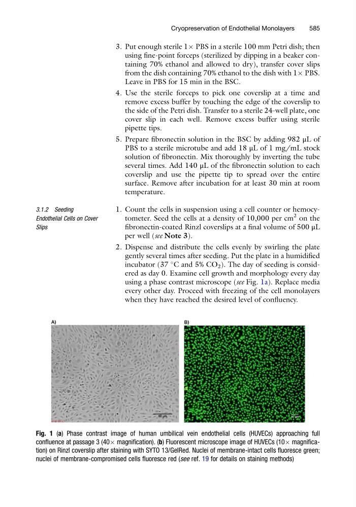
using a phase contrast microscope (see Fig. 1a). Replace media
every other day. Proceed with freezing of the cell monolayers
when they have reached the desired level of confluency.
Fig. 1 (a) Phase contrast image of human umbilical vein endothelial cells (HUVECs) approaching full
confluence at passage 3 (40Â magnification). (b) Fluorescent microscope image of HUVECs (10Â magnification) on Rinzl coverslip after staining with SYTO 13/GelRed. Nuclei of membrane-intact cells fluoresce green;
nuclei of membrane-compromised cells fluoresce red (see ref. 19 for details on staining methods)
Cryopreservation of Endothelial Monolayers
585
using fine-point forceps (sterilized by dipping in a beaker containing 70% ethanol and allowed to dry), transfer cover slips
from the dish containing 70% ethanol to the dish with 1Â PBS.
Leave in PBS for 15 min in the BSC.
4. Use the sterile forceps to pick one coverslip at a time and
remove excess buffer by touching the edge of the coverslip to
the side of the Petri dish. Transfer to a sterile 24-well plate, one
cover slip in each well. Remove excess buffer using sterile
pipette tips.
5. Prepare fibronectin solution in the BSC by adding 982 μL of
PBS to a sterile microtube and add 18 μL of 1 mg/mL stock
solution of fibronectin. Mix thoroughly by inverting the tube
several times. Add 140 μL of the fibronectin solution to each
coverslip and use the pipette tip to spread over the entire
surface. Remove after incubation for at least 30 min at room
temperature.
3.1.2 Seeding
Endothelial Cells on Cover
Slips
1. Count the cells in suspension using a cell counter or hemocytometer. Seed the cells at a density of 10,000 per cm
2 on the
fibronectin-coated Rinzl coverslips at a final volume of 500 μL
per well (see Note 3).
2. Dispense and distribute the cells evenly by swirling the plate
gently several times after seeding. Put the plate in a humidified
incubator (37
C and 5% CO 2 ). The day of seeding is considered as day 0. Examine cell growth and morphology every day
using a phase contrast microscope (see Fig. 1a). Replace media
every other day. Proceed with freezing of the cell monolayers
when they have reached the desired level of confluency.
Fig. 1 (a) Phase contrast image of human umbilical vein endothelial cells (HUVECs) approaching full
confluence at passage 3 (40Â magnification). (b) Fluorescent microscope image of HUVECs (10Â magnification) on Rinzl coverslip after staining with SYTO 13/GelRed. Nuclei of membrane-intact cells fluoresce green;
nuclei of membrane-compromised cells fluoresce red (see ref. 19 for details on staining methods)
Cryopreservation of Endothelial Monolayers
585