

4. Prepare a cell counting chamber with cover glass.
5. Resuspend MK cell suspension, take 10 μL, and mix with 10 μL
of trypan blue in a tube.
6. Fill a counting chamber with 10 μL mixture of cell suspension
and trypan blue.
7. Count cells stained with trypan blue as well as non-stained
(10Â magnification, phase-contrast microscope). The cells
that appear completely blue have damaged membranes that
allow trypan blue to penetrate inside the cell. Such cells are
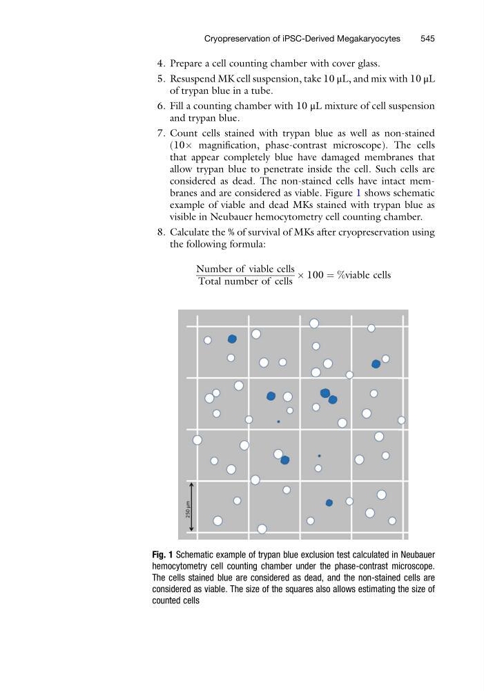
considered as dead. The non-stained cells have intact membranes and are considered as viable. Figure 1 shows schematic
example of viable and dead MKs stained with trypan blue as
visible in Neubauer hemocytometry cell counting chamber.
8. Calculate the % of survival of MKs after cryopreservation using
the following formula:
Number of viable cells
Total number of cells
 100 ¼ %viable cells
Fig. 1 Schematic example of trypan blue exclusion test calculated in Neubauer
hemocytometry cell counting chamber under the phase-contrast microscope.
The cells stained blue are considered as dead, and the non-stained cells are
considered as viable. The size of the squares also allows estimating the size of
counted cells
Cryopreservation of iPSC-Derived Megakaryocytes
545
5. Resuspend MK cell suspension, take 10 μL, and mix with 10 μL
of trypan blue in a tube.
6. Fill a counting chamber with 10 μL mixture of cell suspension
and trypan blue.
7. Count cells stained with trypan blue as well as non-stained
(10Â magnification, phase-contrast microscope). The cells
that appear completely blue have damaged membranes that
allow trypan blue to penetrate inside the cell. Such cells are
considered as dead. The non-stained cells have intact membranes and are considered as viable. Figure 1 shows schematic
example of viable and dead MKs stained with trypan blue as
visible in Neubauer hemocytometry cell counting chamber.
8. Calculate the % of survival of MKs after cryopreservation using
the following formula:
Number of viable cells
Total number of cells
 100 ¼ %viable cells
Fig. 1 Schematic example of trypan blue exclusion test calculated in Neubauer
hemocytometry cell counting chamber under the phase-contrast microscope.
The cells stained blue are considered as dead, and the non-stained cells are
considered as viable. The size of the squares also allows estimating the size of
counted cells
Cryopreservation of iPSC-Derived Megakaryocytes
545