

inside the recipient bursa, trim it to fit by using spring-type
micro-scissors under the microscope.
11. Locate one edge of the ovarian bursa incision, and secure the
transplanted OT by suturing the edge of the bursa to the fat
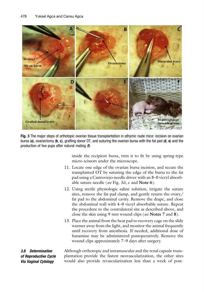
pad using a Castroviejo needle driver with an 8–0 vicryl absorbable suture needle (see Fig. 3d, e and Note 6).
12. Using sterile physiologic saline solution, irrigate the suture
sites, remove the fat-pad clamp, and gently return the ovary/
fat pad to the abdominal cavity. Remove the drape, and close
the abdominal wall with 4–0 vicryl absorbable suture. Repeat
the procedure to the contralateral site as described above, and
close the skin using 9 mm wound clips (see Notes 7 and 8).
13. Place the animal from the heat pad to recovery cage on the slide
warmer away from the light, and monitor the animal frequently
until recovery from anesthesia. If needed, additional dose of
banamine may be administered postoperatively. Remove the
wound clips approximately 7–9 days after surgery.
3.8 Determination
of Reproductive Cycle
Via Vaginal Cytology
Although orthotopic and intramuscular and the renal capsule transplantation provide the fastest neovascularization, the other sites
would also provide revascularization less than a week of postA
B
C
D
E
F
Oviduct
Slit on bursa
Discarded ovary
Grafted donor ovary
Ovariectomy
Re-derived pups
from frozen ovary
Fig. 3 The major steps of orthotopic ovarian tissue transplantation in athymic nude mice: incision on ovarian
bursa (a), ovariectomy (b, c), grafting donor OT, and suturing the ovarian bursa with the fat pad (d, e) and the
production of live pups after natural mating (f)
478
Yuksel Agca and Cansu Agca
micro-scissors under the microscope.
11. Locate one edge of the ovarian bursa incision, and secure the
transplanted OT by suturing the edge of the bursa to the fat
pad using a Castroviejo needle driver with an 8–0 vicryl absorbable suture needle (see Fig. 3d, e and Note 6).
12. Using sterile physiologic saline solution, irrigate the suture
sites, remove the fat-pad clamp, and gently return the ovary/
fat pad to the abdominal cavity. Remove the drape, and close
the abdominal wall with 4–0 vicryl absorbable suture. Repeat
the procedure to the contralateral site as described above, and
close the skin using 9 mm wound clips (see Notes 7 and 8).
13. Place the animal from the heat pad to recovery cage on the slide
warmer away from the light, and monitor the animal frequently
until recovery from anesthesia. If needed, additional dose of
banamine may be administered postoperatively. Remove the
wound clips approximately 7–9 days after surgery.
3.8 Determination
of Reproductive Cycle
Via Vaginal Cytology
Although orthotopic and intramuscular and the renal capsule transplantation provide the fastest neovascularization, the other sites
would also provide revascularization less than a week of postA
B
C
D
E
F
Oviduct
Slit on bursa
Discarded ovary
Grafted donor ovary
Ovariectomy
Re-derived pups
from frozen ovary
Fig. 3 The major steps of orthotopic ovarian tissue transplantation in athymic nude mice: incision on ovarian
bursa (a), ovariectomy (b, c), grafting donor OT, and suturing the ovarian bursa with the fat pad (d, e) and the
production of live pups after natural mating (f)
478
Yuksel Agca and Cansu Agca