

1. Prepare a 5 mg/mL base solution of FDA with acetone in a
glass or polypropylene vessel (such as the Corning 430488
cryo-vial).
2. Prepare a 2.5 μg/mL working solution of FDA by the dilution
of the base FDA solution 2000 times in a phenol red-free
oocyte handling medium (i.e., 1–1999 μL) pre-warmed to
38
C (see Note 21).
3. Incubate groups of oocytes in 500 μL aliquots of the FDA
working solution in 4-well dishes for 2 min at 38
C in dark.
4. Wash oocyte groups twice in 500 μL aliquots of a phenol
red-free oocyte handling medium at 38
C in dark.
5. Observe oocytes under a fluorescence microscope using an
excitation wavelength of 480 nm and emission around
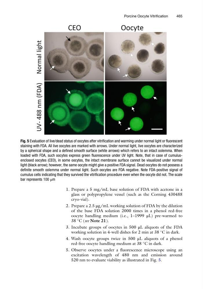
520 nm to evaluate viability as illustrated in Fig. 5.
Fig. 5 Evaluation of live/dead status of oocytes after vitrification and warming under normal light or fluorescent
staining with FDA. All live oocytes are marked with arrows. Under normal light, live oocytes are characterized
by a spherical shape and a defined smooth surface (white arrows) which refers to an intact oolemma. When
loaded with FDA, such oocytes express green fluorescence under UV light. Note, that in case of cumulusenclosed oocytes (CEO), in some oocytes, the intact membrane surface cannot be visualized under normal
light (black arrow); however, the same oocyte might give a positive FDA signal. Dead oocytes do not possess a
definite smooth oolemma under normal light. Such oocytes are FDA negative. Note FDA-positive signal of
cumulus cells indicating that they survived the vitrification procedure even when the oocyte did not. The scale
bar represents 100 μm
Porcine Oocyte Vitrification
465
glass or polypropylene vessel (such as the Corning 430488
cryo-vial).
2. Prepare a 2.5 μg/mL working solution of FDA by the dilution
of the base FDA solution 2000 times in a phenol red-free
oocyte handling medium (i.e., 1–1999 μL) pre-warmed to
38
C (see Note 21).
3. Incubate groups of oocytes in 500 μL aliquots of the FDA
working solution in 4-well dishes for 2 min at 38
C in dark.
4. Wash oocyte groups twice in 500 μL aliquots of a phenol
red-free oocyte handling medium at 38
C in dark.
5. Observe oocytes under a fluorescence microscope using an
excitation wavelength of 480 nm and emission around
520 nm to evaluate viability as illustrated in Fig. 5.
Fig. 5 Evaluation of live/dead status of oocytes after vitrification and warming under normal light or fluorescent
staining with FDA. All live oocytes are marked with arrows. Under normal light, live oocytes are characterized
by a spherical shape and a defined smooth surface (white arrows) which refers to an intact oolemma. When
loaded with FDA, such oocytes express green fluorescence under UV light. Note, that in case of cumulusenclosed oocytes (CEO), in some oocytes, the intact membrane surface cannot be visualized under normal
light (black arrow); however, the same oocyte might give a positive FDA signal. Dead oocytes do not possess a
definite smooth oolemma under normal light. Such oocytes are FDA negative. Note FDA-positive signal of
cumulus cells indicating that they survived the vitrification procedure even when the oocyte did not. The scale
bar represents 100 μm
Porcine Oocyte Vitrification
465