

2.3 Tools
for Warming Vitrified
Samples
1. Stereo microscope to visualize oocytes/zygotes during
handling (recommended: Olympus SZX16) equipped with a
warmed stage or a portable warm plate (such as Microwarm
Plate MP-1000, Kitazato, Japan).
2. A stand-alone warm plate (such as SP-45D, Hirasawa, Tokyo).
3. A 35 mm Petri dish (Falcon 351008) and a 4-well dish (Nunc
MultiDishes, Thermo Fisher Scientific) for the warming and
stepwise re-hydration, respectively of each oocyte lot.
4. Fine glass pipettes with a mouth piece, as described above
(in Subheading 2.2). Two pipettes are required: (a) to move
oocytes/zygotes from the warming dish to-and-through the
de-hydration dish, (b) to move oocytes/zygotes from the
de-hydration dish to the post-warming.
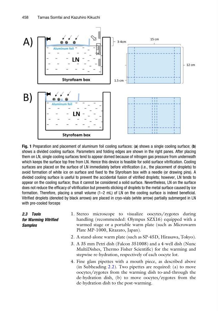
Fig. 1 Preparation and placement of aluminum foil cooling surfaces: (a) shows a single cooling surface; (b)
shows a divided cooling surface. Parameters and folding edges are shown in the right panes. After placing
them on LN, single cooling surfaces tend to appear domed because of nitrogen gas pressure from underneath
which keeps the surface top free from LN. Hence this device is feasible for solid surface vitrification. Cooling
surfaces are placed on the surface of LN immediately before vitrification (i.e., the placement of droplets) to
avoid formation of white ice on surface and fixed to the Styrofoam box with a needle (or drawing pins). A
divided cooling surface is useful to prevent the accidental fusion of vitrified droplets; however, LN tends to
appear on the cooling surface; thus it cannot be considered a solid surface. Nevertheless, LN on the surface
does not reduce the efficacy of vitrification but prevents sticking of droplets to the metal surface caused by ice
formation. Therefore, placing a small volume (1–2 mL) of LN on the cooling surface is indeed beneficial.
Vitrified droplets (denoted by black arrows) are placed in cryo-vials (white arrow) partially submerged in LN
with pre-cooled forceps
458
Tamas Somfai and Kazuhiro Kikuchi
for Warming Vitrified
Samples
1. Stereo microscope to visualize oocytes/zygotes during
handling (recommended: Olympus SZX16) equipped with a
warmed stage or a portable warm plate (such as Microwarm
Plate MP-1000, Kitazato, Japan).
2. A stand-alone warm plate (such as SP-45D, Hirasawa, Tokyo).
3. A 35 mm Petri dish (Falcon 351008) and a 4-well dish (Nunc
MultiDishes, Thermo Fisher Scientific) for the warming and
stepwise re-hydration, respectively of each oocyte lot.
4. Fine glass pipettes with a mouth piece, as described above
(in Subheading 2.2). Two pipettes are required: (a) to move
oocytes/zygotes from the warming dish to-and-through the
de-hydration dish, (b) to move oocytes/zygotes from the
de-hydration dish to the post-warming.
Fig. 1 Preparation and placement of aluminum foil cooling surfaces: (a) shows a single cooling surface; (b)
shows a divided cooling surface. Parameters and folding edges are shown in the right panes. After placing
them on LN, single cooling surfaces tend to appear domed because of nitrogen gas pressure from underneath
which keeps the surface top free from LN. Hence this device is feasible for solid surface vitrification. Cooling
surfaces are placed on the surface of LN immediately before vitrification (i.e., the placement of droplets) to
avoid formation of white ice on surface and fixed to the Styrofoam box with a needle (or drawing pins). A
divided cooling surface is useful to prevent the accidental fusion of vitrified droplets; however, LN tends to
appear on the cooling surface; thus it cannot be considered a solid surface. Nevertheless, LN on the surface
does not reduce the efficacy of vitrification but prevents sticking of droplets to the metal surface caused by ice
formation. Therefore, placing a small volume (1–2 mL) of LN on the cooling surface is indeed beneficial.
Vitrified droplets (denoted by black arrows) are placed in cryo-vials (white arrow) partially submerged in LN
with pre-cooled forceps
458
Tamas Somfai and Kazuhiro Kikuchi